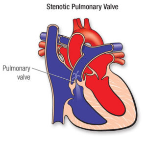
Pulmonary valve stenosis

Pulmonary valve stenosis refers to an obstruction of the valve that is located within the upper right chamber of the heart (right ventricle) and the lung arteries (pulmonary arteries). When a heart valve is narrowed the valve’s flaps (cusps) could become stiff or stiff. This can reduce heart valve blood flow¹.
Typically, pulmonary valve dysfunction is a result of a heart issue that occurs prior to birth (congenital heart defects). However, adult patients may experience pulmonary valve stenosis due to an outcome of another condition.
The severity of pulmonary valve stenosis varies from moderate to severe. A few people suffering from moderate pulmonary valve stenosis may not have any symptoms and might just require periodic doctor’s visits. A severe and moderate pulmonary valve stenosis could require a procedure to fix and replace the valve.
Symptoms
The symptoms and signs vary according to the degree of blood flow is affected. A few people suffering from mild pulmonary stenosis do not show symptoms. People with more severe pulmonary stenosis might first experience symptoms when exercising.
Signs and symptoms could be:
- A loud sound (murmur), which can be heard by a microphone
- Fatigue
- Breathlessness, particularly during exercise
- Chest pain
- The loss of awareness (fainting)²
Babies born with pulmonary valve stenosis as well as other heart defects that are congenital may be to be blue (cyanotic).
When to seek medical help?
Discuss with your doctor If either of your children suffers from:
- Breathing problems
- Chest pain
- Fainting
Should you suspect that your kid is suffering from pulmonary stenosis, or another heart issue timely diagnosis and treatment may lower the chance of complications.
Causes
Pulmonary valve stenosis is often caused by a heart defect that is congenital. The cause of the problem is not clear. The pulmonary valve does not develop in a proper way when the baby grows inside the womb.
The pulmonary valve is comprised of three small pieces of tissue, known as flaps (cusps). The cusps can be opened and closed at every heartbeat, and help ensure that blood flows in the correct direction.
In the case of pulmonary valve stenosis one or more cusps could be thick or stiff or might connect in fusion. This means that the valve isn’t fully open. The valve’s smaller opening makes it difficult for blood to drain out of the lower chamber of the heart (right ventricle). Pressure rises inside the right ventricle when it tries to pump blood through the narrower opening. The pressure increase causes an increase in heart strain that ultimately results in the right ventricle’s muscular wall getting thicker.
Risk factors
Conditions or diseases that could raise the chance of developing pulmonary valve stenosis are:
- German measles (rubella). Having German measles (rubella) during pregnancy can increase the chance of developing pulmonary valve stenosis for the infant.
- Noonan syndrome. This genetic disorder can cause a variety of problems to the heart’s structure as well as function.
- Rheumatic fever. This complication of strep throat could cause permanent heart damage and valves of the heart. It may increase the chance to develop pulmonary valve stenosis in the course of.
- Carcinoid Syndrome. A rare cancerous tumor releases chemicals into bloodstreams which can cause breathlessness flushing, as well as other symptoms and signs. A few people who suffer from this syndrome develop heart diseases called carcinoid, which causes damage to heart valves.
Complications
Potential complications of pulmonary narrowing include:
- An infection of the inner lining of the heart (infective endocarditis). People with heart valve issues such as pulmonary stenosis, face an increased chance of developing bacteria-related infections which affect the heart’s lining.
- A heartbeat that is irregular (arrhythmia). People who suffer from pulmonary stenosis are more likely to experience an unsteady heartbeat. If the stenosis isn’t serious, irregular heartbeats caused by pulmonary stenosis typically aren’t serious.
- The heart muscle is weakened by the muscles. In severe pulmonary stenosis, the right ventricle has to pump harder to push the circulation of blood through the blood vessel. The stress upon the heart leads to the muscle walls of the ventricle to become thicker (right hypertrophy of the ventricular wall).
- heart failure. If the right ventricle isn’t pumping properly then heart failure will develop. Heart failure symptoms include fatigue, breathlessness along with swelling and pain in the abdomen and legs.
- The complications of pregnancy. The risks of complications during labor and birth are greater for women with severe pulmonary valve stenosis compared to those who don’t have the condition.
Diagnosis
The condition is typically detected in the early years of childhood. However, it could not be discovered until later in life.
The doctor will utilize the stethoscope for listening to your child’s or your child’s heart. A squealing sound (murmur) is caused by the choppy (turbulent) circulation of blood over the valve could be heard.
The tests to determine if you have pulmonary valve stenosis can consist of:
- Electrocardiogram (ECG or EKG). This simple and painless test detects electrical signals within the heart. These sticky patches (electrodes) are put on the chest and often the legs and arms. The electrodes are connected by wires to a computer that shows the results of the test. An ECG It can reveal how the heart beats. It could identify the signs of thickening of the heart muscle.
- Echocardiogram. An echocardiogram uses sound waves to create photographs of the heart. This type of test permits a physician to observe what the heart does as it pumps blood. An echocardiogram will reveal the shape of the pulmonary valve as well as the extent and location of any narrowing of the valve.
- Catheterization of the heart. A thin tube (catheter) is placed into the groin and then threaded through blood vessels that lead to the heart. The dye can be injected via the catheter into blood vessels in order to make them apparent in an X-ray (coronary angiogram).Doctors also employ cardiac catheterization to gauge pressure inside those chambers in the heart, to assess the force with which blood flows into the heart. When you’ve had a diagnosis of pulmonary valve stenosis your doctor can tell the severity of the problem by comparing the variation on blood pressures between the left lower chamber of your heart and the pulmonary arterial.
- Other tests for imaging. Magnetic resonance imaging (MRI) and computed tomography (CT) scans can be employed to determine the severity of the pulmonary valve stenosis.
Treatment
If you suffer from moderate pulmonary valve stenosis that does not cause symptoms, you may just require regular doctor’s checks.
If you suffer from severe or moderate pulmonary valve narrowing you might require an operation on your heart or surgery. The kind of procedure or procedure you undergo will depend on the overall condition of your body as well as the appearance of the pulmonary valve.
Heart surgeries and procedures used in treating the condition of pulmonary valve stenosis are:
- Balloon valvuloplasty. The doctor inserts an elastic tubing (catheter) with balloons on its end into an artery, typically in the groin. X-rays are employed to guide the catheter towards the valve that has narrowed inside the heart. The doctor fills this balloon. It expands the valve’s opening and also separates the valve flaps when required. After that, the balloon deflates. The balloon and catheter are then removed.Valvuloplasty can enhance blood flow through the heart and decrease the symptoms of pulmonary valve stenosis. However, the valve could shrink once more. Certain people require valve replacement or repair in the near future.
- Replacement of the pulmonary valve. If balloon valvuloplasty isn’t feasible, an open-heart surgical procedure or even a catheter could be performed to repair the valve in the pulmonary. If there are any other congenital heart problems The doctor will usually treat them in the same procedure.People who have undergone the replacement of their pulmonary valves must have antibiotics taken prior to specific dental treatments or surgeries in order to avoid endocarditis.
Lifestyle and home solutions for home and lifestyle
If you suffer from valve disease it is important to ensure that you are keeping your heart in good health. Certain lifestyle choices can lower the chance of developing different types of heart disease or even having heart attacks.
Changes in your lifestyle that you should talk to your doctor might include:
- Quitting smoking
- A heart-healthy diet includes vegetables fruits and dairy products with low-fat Whole grains, lean meat
- Weight maintenance is essential to maintain a healthy weight
- Getting regular exercise

Good day! Do you know if they make any plugins to
help with SEO? I’m trying to get my site to rank for some targeted keywords but I’m not seeing very good success.
If you know of any please share. Kudos! You can read similar art here: Wool product
Hi there! Do you know if they make any plugins to assist with SEO?
I’m trying to get my site to rank for some targeted keywords but I’m not seeing very good
gains. If you know of any please share. Thanks!
You can read similar article here: Your destiny
genting casino southend
References:
http://wudao28.com/home.php?mod=space&uid=2234296
hard rock casino punta cana
References:
https://escatter11.fullerton.edu/nfs/show_user.php?userid=9406715
jackpot capital casino
References:
https://www.demilked.com/author/callnerve76/
laughlin casinos
References:
https://www.google.com.co/url?q=https://hedge.fachschaft.informatik.uni-kl.de/s/–P1p-ub5
casino charlevoix
References:
https://www.google.dm/url?q=https://www.udrpsearch.com/user/iconwall12
blackjack mountain oklahoma
References:
http://taikwu.com.tw/dsz/home.php?mod=space&uid=3089584
casino aztar evansville
References:
https://www.orkhonschool.edu.mn/profile/larsonbochenneberg78562/profile
merkur online casino
References:
https://www.holycrossconvent.edu.na/profile/lohmannjyjcoughlin76152/profile