
Diagnosis
Key Diagnostic points:
- The usual presentation is systemic hypertension.
- Echocardiography/Doppler is diagnostic; a peak gradient of more than 20 mm Hg may be significant due to collaterals around the coarctation reducing gradient despite the severe obstruction.
- The bicuspid aortic valve is found in between 50 and 80 percent of patients.
- A delayed pulse in the femoral artery as compared to brachial the artery.
- Systolic pressures are higher in the upper extremities but lower in the lower extremities. Diastolic pressures are comparable.
The age at when the coarctation of the aorta is recognized is contingent upon what severity is associated with the issue. If the coarctation of the aorta is serious, it’s usually detected at the time of infancy. Coarctation of the aorta can be often detected on sonograms of the fetus.
- Adults and children older than age 10 who have been diagnosed with coarctation of the Aorta can be suffering from milder cases but are not afflicted with symptoms. They are often healthy until the doctor determines:
- Blood pressure that is high in the arms
- A difference in blood pressure between legs and arms is characterized by more blood pressure in arms, and lower blood pressure in the legs.
- A slow or weak pulse in the legs.
- A heart murmur – an unusual whooshing sound caused by a faster flow of blood through the narrowed region
Tests
Diagnostic tests for confirmation that coarctation is present in the aorta could be:
Echocardiogram. Echocardiograms use sound waves to create pictures of the heart which can be watched on a screen. The test will often reveal to your doctor the location and the severity of the coarctation of the aorta. It could also reveal other heart problems like an aortic valve that is bicuspid. Echocardiograms are often used by doctors to detect coarctation of the Aorta and to determine the best treatments for you.
Electrocardiogram (ECG). An electrocardiogram monitors the electrical signals generated by your heart. In this test the patches are sticky (electrodes) are connected to your chest and the limbs. They are connected by wires that connect to an electronic monitor. They record electrical signals that cause your heart to beat. Computers record the data and display it in waves on a display as well as on paper.
If the coarctation Aorta is extreme The ECG could show thickening of the wall of the lower chambers (ventricular hypertrophy).
Chest Xray. A chest X-ray produces images of your lungs and heart. A chest X-ray may reveal a narrowing of the aorta near the point of coarctation, or an expanded portion of the aorta both.
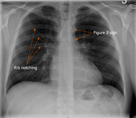
Radiography can reveal scalloping of the lower portion of the ribs (rib notching) due to the expansion of the collateral intercostal vessels. The left subclavian arterial artery and dilation of the aortic poststenotically, as well as LV swelling, could be present. The coarctation region, as well as the post stenotic dilation of ascending aorta, can cause a “3” sign along with the shadow of the aortic region appearing on the PA chest radiograph (the hole inside”3″ is the “3” representing the area of coarctation).
Magnetic resonance imaging (MRI). An MRI utilizes a powerful magnetic field as well as radio waves to provide precise images of the blood vessels and the heart. The test will reveal the exact location and severity of coarctation of the aorta, as well as damage to blood vessels in other areas, as well as any other heart problems. Your physician may also utilize MRI results to determine the best treatment.
The computerized (CT) scan. A CT scan utilizes a series of X-rays in order to produce detailed cross-sectional pictures of the body.
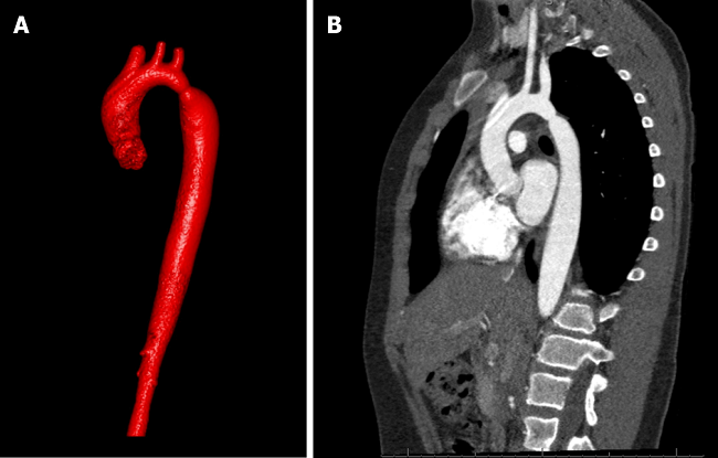
CT Angiogram. A CT angiogram utilizes a dye and images to reveal the interior of the coronary arteries. It shows the flow of blood in the veins as well as the arteries. The test will reveal the extent and location of the coarctation of your aorta as well as determine if it has an effect on other blood vessels throughout your body. A CT angiogram may also be used to determine other heart problems or determine the best treatment options.
Catheterization of the cardiac. During this procedure, the doctor places an extremely thin, long tubing (catheter) in an artery, vein or inside your arm, groin or neck, then connects it into your heart via X-ray imaging. Sometimes dye is injected through the catheter to make your heart’s organs show more clearly on images from X-rays.
Cardiac catheterization is a way to identify the degree of coarctation of the aortic artery. The doctor might use it to aid in planning surgical procedures or other treatments should you require it. Catheter procedures could also be utilized to carry out certain procedures for coarctation of the Aorta.
Treatment
Treating coarctation on the aorta is based on your age at the moment of diagnosis and the seriousness of your situation. Other heart problems could be treated in the same way in the event of aortic coarctation.
A specialist in congenital heart diseases will examine you and determine the most suitable treatment for your specific condition.
Medication
It isn’t a treatment for an aorta coarctation. However, your doctor might suggest it lower blood pressure prior to and after the placement of stents or surgeries. Even though repairing the aortic coarctation helps improve blood pressure, many require blood pressure medications following the successful operation or the stenting procedure.
Babies suffering from severe coarctation of their aorta typically receive medicine that keeps the arteriosus ductus open. This allows blood to circulate through the constriction until coarctation heals.
The procedure of surgery or any other
There are many surgeries that can be performed to treat aortic coarctation. Your physician can inform you of the type of surgery that is most likely to repair the condition of your child or yourself. There are a variety of options to choose from:
Resection using an anastomosis from end to end. This method involves the removal of the narrowed portion of the aorta (resection) then joining the two healthy sections of the aorta (anastomosis).
Aortic flap subclavian. A part of the blood vessel that supplies blood to the left arm (left subclavian arterial) could be utilized to increase the area that is narrowed by the aorta.
Bypass repair of grafts. This technique involves bypassing the narrowed region by inserting a tube known as a graft between two parts that make up the aorta.
Patch Aortoplasty. Your doctor might treat the coarctation by cutting through the narrowed region of the aorta before attaching a patch made of synthetic material that will widen the blood vessels. Patch angioplasty can be beneficial in cases where the coarctation is long segments in the aorta.
Stenting and balloon angioplasty
The procedure is often used as a primary treatment for aortic coarctation, instead of surgery. It could be performed if the narrowing happens following coarctation surgery.
In balloon angioplasty, the doctor inserts an extremely thin elastic tubing (catheter) in an artery that runs through the groin area and threads it through the blood vessels to the heart via an X-ray image.
The doctor will place an uninflated balloon in the opening in the narrowed Aorta. Once it is inflated, your aorta opens and blood flow becomes easier. In some cases, the hollow tube is covered in mesh (stent) is put in the aorta to keep narrowed Aorta open.