Coarctation of the Aorta: Symptoms, Treatments & Tests
Overview of Coarctation of the aorta
The aorta is the longest arterial system within your body. It carries rich oxygenated blood to your entire body. Aorta narrowing (aortic coarctation) makes your heart pump more forcefully to transport oxygen-rich blood around the aorta.
Aortic Coarctation is generally present at birth (congenital). Although it can be affecting any part of the aorta the problem is usually situated in the vicinity of a blood vessel, known as the ductus arteriosis. The symptoms can be minor to serious. It is possible that it will not be noticed until later in life, based on how far the aorta has narrowed.
The coarctation of the aorta is often occurred and is found in conjunction with other heart issues. Although treatment can be successful, this condition requires regular monitoring for the rest of your life.
Symptoms
Aortic coarctation symptoms are triggered by the intensity of the problem. Many people don’t show symptoms. Mild coarctation might not be detected until the age of adulthood.
Babies suffering from severe coarctation of their aorta could start experiencing symptoms soon after the birth. This includes:
Pale skin
Trouble breathing
Irritability
Heavy sweating
Food insecurity
People who suffer from coarctation of the Aorta might also exhibit signs or symptoms that suggest other heart issues that are often associated in conjunction with the condition.
A sign or symptom for coarctation in the aorta that occurs after infancy typically include:
Get medical attention If you or your child is suffering from any of the following symptoms or signs:
Severe chest pain
Fainting
Shortness of breath that is sudden
Unexpectedly high blood pressure
Although having these symptoms or signs doesn’t necessarily mean you’re suffering from an illness of a serious nature It is recommended to be examined immediately. Early detection and treatment may be the difference between life and death.
Causes
The etiology of aortic coarctation is not well established. It is usually present from birth (congenital). Congenital heart defects are among the most prevalent of birth defects.
Sometimes, coarctation of the aorta is seen in later years of life. The causes or circumstances that may make the aorta narrow and lead to this problem are:
Traumatic injury
Acute hardening of the arteries (atherosclerosis)
Inflamed arterial arteries (Takayasu’s arteritis)
The coarctation of the aorta typically occurs in the blood vessels which branch out towards your upper body, and also before the blood vessels which connect to the lower part of your body. It can lead to elevated blood pressure within your arm, but lower levels of blood pressure within your ankles and legs.
In the event of coarctation of the aorta, the lower left chamber (left ventricle) of your heart functions more difficult in order to move blood around the narrowed aorta and the blood pressure rises within the left ventricle. This can result in the wall of the left ventricle becoming thicker (hypertrophy).
Risk factors
Coarctation of the aorta typically is associated with other heart defects that are congenital. Certain heart diseases are frequently related to coarctation, such as:
Bicuspid Aortic Valve. The aortic valve divides the lower left chamber (left ventricle) of the heart from the aorta. A bicuspid aortic valve is made up of 2 flaps (cusps) in place of the normal three. A large number of patients with coarctation of their aortas have an aortic valve that is bicuspid.
The condition of sub-aortic stenosis. Sub-aortic stenosis occurs when there is a narrowing in the region beneath the aortic valve, which blocks blood from the left ventricle towards the Aorta. This narrowing can take an amorphous fibrous membrane.
Patent ductus Arteriosus. The ductus arteriosus is an arterial blood vessel that connects the left pulmonary artery of a baby and the aorta. It lets blood flow through the lungs when the baby is developing during the womb. Soon after birth the ductus arteriosus normally closes. If it’s still open, it’s known as an arteriosus patent ductus.
Holes in the walls connect the right and left parts of your heart. Some people are born with an opening within the heart’s walls (septum) that connects the upper chambers of the heart (atrial septal defect) or between the lower chambers (ventricular septal defect). This creates oxygen-rich blood on the left heart side of it to blend with blood that is oxygen-poor on the right side of the heart.
Mitral valve stenosis is congenital. The mitral valve is located between the lower and upper left chambers of your heart. It allows blood flow to on the left-hand side of your heart. Mitral valve stenosis is when the valve narrows. In a result, blood circulation between the lower and upper left chambers of the heart is reduced and the pressure is increased in the left upper chamber (atrium). The oxygen-rich blood that comes from the lungs flows back to the heart via veins that join to the left upper chamber. A rise in blood pressure within the left atrium can cause symptoms of lungs congestion. The symptoms may include breathing difficulty, shortness of breath breathing during exercise, and shortness of breath while lying down.
The aorta’s coarctation is more prevalent for males than females, as well for those suffering from certain genetic conditions like Turner syndrome.
Complications
If not treated coarctation of the aorta often results in complications. In infants, it could result in heart failure or even death.
Blood pressure that is high is the most frequently reported long-term result from coarctation within the Aorta. The blood pressure typically decreases after the aortic coarctation is corrected; however, it can be more than normal.
Other complications associated with coarctation of the Aorta can be:
A bulging or weak artery inside the cerebral artery (brain aneurysm) or bleeding inside the cerebral area (hemorrhage)
Aortic tear or rupture (dissection)
An increase in the size of a portion from the wall that forms the aorta (aneurysm)
Heart failure
Premature coronary artery diseases -an increase in blood vessels that provide the heart
Stroke
If the coarctation of your aorta is serious the heart may not be able enough to pump blood to the other organs. This could result in heart damage and could cause kidney failure or other organ problems.
There are complications that can occur following treatment for coarctation of the aorta. They can include:
Aorta narrowing (re-coarctation possible several years after treatment)
High blood pressure
Aortic aneurysm, or rupture
You’ll require a lifetime examination for the coarctation of your aorta. Additionally, you could require further treatments
Prevention
Aortic coarctation cannot be prevented since it’s typically present from the time of birth. But, if your child is suffering from an issue that can increase the risk of developing aortic coarctation, like Turner syndrome, bicuspid aortic valve, or other cardiovascular defect or has a family history of congenital heart diseases early detection may assist. Discuss the possibility of coarctation of the aortic artery with your physician.
Wow, amazing blog format! How long have you been blogging
for? you make blogging look easy. The total glance of your web site is great, as neatly
as the content material! You can see similar here sklep
Wow, marvelous blog structure! How long have you been running a blog for?
you make blogging look easy. The total look of your
website is excellent, let alone the content! You can see similar here sklep internetowy
Wow, awesome blog layout! How lengthy have you ever been running a blog for?
you make blogging look easy. The whole look of your web site is
excellent, as well as the content! You can see similar here sklep online
Hello just wanted to give you a quick heads up.
The text in your content seem to be running off the screen in Safari.
I’m not sure if this is a format issue or something to do with web
browser compatibility but I figured I’d post to let you know.
The layout look great though! Hope you get the issue resolved soon. Kudos I
saw similar here: Sklep online
Wow, incredible blog format! How long have you been running a
blog for? you make blogging look easy. The whole glance of your website is great, as smartly as the content!
You can see similar here sklep internetowy
It’s very interesting! If you need help, look here: ARA Agency
Hello! Do you know if they make any plugins to assist
with Search Engine Optimization? I’m trying to get my blog to rank for some targeted
keywords but I’m not seeing very good success. If you know
of any please share. Thank you! You can read similar
article here: Sklep internetowy
Hey! Do you know if they make any plugins to help with SEO?
I’m trying to get my blog to rank for some targeted keywords but I’m not seeing very
good results. If you know of any please share. Many thanks!
You can read similar text here: Sklep
Hello there! Do you know if they make any plugins to help with SEO?
I’m trying to get my blog to rank for some targeted keywords but I’m not seeing very good results.
If you know of any please share. Many thanks! You can read similar art
here: Sklep internetowy
Hello! Do you know if they make any plugins to help with Search Engine Optimization? I’m trying to get my
website to rank for some targeted keywords but I’m not seeing very good
gains. If you know of any please share. Thanks!
You can read similar art here: GSA Verified List
Hey! Do you know if they make any plugins to assist with Search Engine Optimization?
I’m trying to get my blog to rank for some targeted keywords but I’m not seeing very good success.
If you know of any please share. Cheers! I saw similar art here: Link Building
That was a great blog. You made some exceptional points and I appreciate for your insight!
very good post, i definitely love this site, go on it
Howdy my name is Kenneth Kennemore and concerning looked each and every method posssible to be able to easy, Concerning concentrated these individuals into just the most effective people who deliever what exactly that they declare. Become a member of my own ZERO COST Cash Points ezine jacksonville divorce attorney plus allow us to provide you with learning to make dollars on the internet fast and easy.
My cousin and I had been debating this topic, he is generally looking to show me wrong. Your view on this is fantastic and exactly how I really think. I just sent him this web site to demonstrate him your point of view. After browsing around your site I book-marked and will be back to read your new posts!
Nice read, I just passed this onto a friend who was doing a little research on that. And he just bought me lunch as I found it for him smile Therefore let me rephrase that: Thanks for lunch!
pretty practical stuff, overall I consider this is worth a bookmark, thanks
This is a great blog, will you be involved in doing an interview about how you developed it? If so e-mail me!
This is an astonishing entry. Thank you very much for the supreme post provided! I was looking for this entry for a long time, but I wasn’t able to find a trustworthy source.
you have got a great blog right here! would you like to make some invite posts on my weblog?
Have you ever thought about publishing an ebook or guest authoring on other websites? I have a blog based upon on the same information you discuss and would really like to have you share some stories/information. I know my audience would value your work. If you are even remotely interested, feel free to send me an email.
i can see lots of free music on the internet but most of them are pirated. *
This can be a enormous plus a highly intriguing e-mail have a look at on this excellent weblog.
I really like seeing websites that understand the value of providing a quality useful resource for free. I wish I had your blogging style.
This is certainly a remarkably amazing powerful resource that you’re offering and you simply provide it away cost-free!! I that can compare with discovering websites ones understand the particular in providing you fantastic learning resource for zero cost. We truly dearly loved examining this web site. Appreciate it!
Interesting blog! Is your theme custom made or did you download it from somewhere? A theme like yours with a few simple tweeks would really make my blog jump out. Please let me know where you got your design. With thanks
It is the best time to make some plans for the future and it is time to be happy. I’ve read this post and if I could I want to suggest you some interesting things or suggestions. Maybe you could write next articles referring to this article. I wish to read more things about it!
my grandmother is always into herbal stuffs and she always say that ayurvedic medicines are the best stuff’
whoa, this is a really good piece of information. I read about something like this before, this is impressively great stuff.
Hi excellent website! Does running a blog similar to this take a great deal of work? I’ve absolutely no expertise in programming but I was hoping to start my own blog in the near future. Anyways, should you have any suggestions or tips for new blog owners please share. I understand this is off topic but I simply needed to ask. Thanks!
Recommeneded websites… […]Here are some of the sites we recommend for our visitors[…]……
i believe,When misery restorative healing, some sort of unhappy restful sample, but even check out the trip if noticeable consumed. Good care of the night time, insert your name installing while in the eyeport, glazing is a love, Wanted Recollection is sweet.
I am impressed with this site, really I am a fan .
What refreshing delight your posts are. I understood this should be here yet I ultimately became fortunate and the search terms worked. Getting more people to be part of the discussion is actually a positive thing.
Very good blog you have here but I was curious if you knew of any user discussion forums that cover the same topics discussed here? I’d really love to be a part of group where I can get opinions from other knowledgeable individuals that share the same interest. If you have any suggestions, please let me know. Many thanks!
Great website you got here! Yoo man great reads, post some more! Im gon come back so better have updated
Have you ever considered creating an e-book or guest authoring on other websites? I have a blog based on the same topics you discuss and would love to have you share some stories/information. I know my readers would enjoy your work. If you’re even remotely interested, feel free to shoot me an e mail.
Howdy! Do you know if they make any plugins to help with Search Engine Optimization? I’m trying to get my blog to rank for some
targeted keywords but I’m not seeing very good results. If you know of any please share.
I would like to thnkx for the efforts you’ve put in writing this website. I am hoping the same high-grade blog post from you in the upcoming as well. In fact your creative writing skills has encouraged me to get my own web site now. Really the blogging is spreading its wings rapidly. Your write up is a good example of it.
My wife and i ended up being absolutely delighted Louis managed to carry out his research with the precious recommendations he discovered while using the web page. It is now and again perplexing just to choose to be giving for free methods which often most people have been selling. We understand we need the writer to thank for that. Those explanations you made, the simple site menu, the relationships you will help to foster – it is mostly exceptional, and it’s really aiding our son in addition to the family do think that issue is entertaining, which is extremely important. Many thanks for all!
Hey there just wanted to give you a quick heads up. The words in your article seem to be running off the screen in Safari. I’m not sure if this is a format issue or something to do with web browser compatibility but I figured I’d post to let you know. The style and design look great though! Hope you get the problem solved soon. Thanks
Great website. You’ll discover several opinions on this matter and also this blog declares the matter quite fantastic.
there is a need for firming lotion so that we can always maintain the health of our skin ,
very good post, i definitely love this website, keep on it
After study a number of the websites with your site now, and that i genuinely appreciate your method of blogging. I bookmarked it to my bookmark site list and are checking back soon. Pls have a look at my web page likewise and let me know if you agree.
I am typically to blogging and that i really appreciate your site content. This great article has really peaks my interest. My goal is to bookmark your site and maintain checking for brand spanking new data.
I discovered your blog site internet site on bing and check a couple of your early posts. Always maintain the good operate. I simply extra the Feed to my MSN News Reader. Looking for toward reading a lot more from you at a later time!…
Hi. best wishes to you and your very nice blog,
Aw, it was an extremely nice post. In thought I have to invest writing such as this moreover – spending time and actual effort to produce a very good article… but what can I say… I procrastinate alot and also no indicates find a way to get something accomplished.
certainly like your web site but you have to take a look at the spelling on quite a few of your posts. Several of them are rife with spelling problems and I in finding it very bothersome to inform the reality then again I will surely come again again.
Wonderful write-up, I have book marked this web-site. I wish I had your insight.
Pretty! This was an incredibly wonderful article. Many thanks for supplying these details.
The my fantastic satisfaction to consider your site also to appreciate your own outstanding submit right here. I prefer which greatly. I am aware that you put a lot attention for those articles, as these make sense and are very useful
An interesting discussion may be worth comment. There’s no doubt that you should write on this topic, it will not be a taboo subject but generally folks are inadequate to speak on such topics. Yet another. Cheers
Hey this is a good post. I’m going to email this to my friends. I stumbled on this while browsing on aol I’ll be sure to come back. thanks for sharing.
Your website is beautifully decorated and effortlessly navigated. We have enjoyed going to this internet site currently and hope to go to quite a few additional occasions inside future.
i love to collect different models of cellphones that is why i have lots of cellphones at home.,
Good write-up, I¡¦m regular visitor of one¡¦s site, maintain up the nice operate, and It is going to be a regular visitor for a lengthy time. Pristina Travel
I’ve been browsing online more than three hours as of late, but I never found any interesting article like yours. It is lovely worth enough for me. Personally, if all webmasters and bloggers made just right content as you probably did, the net will be a lot more helpful than ever before!
You have good command on this topic and have explained in a very pleasant way. Thanks for sharing.
Hey, have you ever before wondered to write regarding Nintendo or PS handheld?
i get my art books from barnes and noble that come or amazon, they have some great discount coupons too.,
There is some validity but I will hold judgment until I look into it further
After study a number of of the blog posts in your web site now, and I actually like your method of blogging. I bookmarked it to my bookmark website record and will be checking again soon. Pls take a look at my website as well and let me know what you think.
Have you already setup a fan page on Facebook ?”`”.’
Do you have a spam issue on this website; I also am a blogger, and I was wondering your situation; many of us have developed some nice practices and we are looking to swap solutions with other folks, please shoot me an e-mail if interested.
I am glad to be one of the visitors on this great site (:, appreciate it for posting.
bathroom towels should be maintained with a good fabric conditioner so that they will last longer::
Seriously this kind of guide is definitely amazing it truly helped me as well as my children, appreciate it!
When I originally commented I clicked the -Notify me when new comments are added- checkbox and now every time a comment is added I get four emails with the same comment. Is there any way you are able to eliminate me from that service? Thanks!
Greetings, May I download your page image and make use of it on my own blog site?
Hi there, I do think your blog might be having internet browser compatibility issues. Whenever I take a look at your site in Safari, it looks fine however when opening in IE, it’s got some overlapping issues. I merely wanted to provide you with a quick heads up! Besides that, wonderful site!
You lost me, friend. I mean, I assume I get what youre indicating. I have an understanding of what you’re saying, but you just appear to have forgotten about that you will find some other people in the world who look at this matter for what it genuinely is and might not agree with you. You may be turning away a decent amount of persons who might have been fans of your blog.
wonderful post, very informative. I’m wondering why the other specialists of this sector don’t understand this. You should continue your writing. I am sure, you have a great readers’ base already! rent a car kosovo
I’m writing to make you know of the fabulous encounter my wife’s child undergone going through the blog. She realized a good number of things, not to mention what it’s like to have an excellent giving spirit to make certain people just learn certain extremely tough subject areas. You undoubtedly surpassed readers’ desires. Many thanks for presenting the useful, trustworthy, revealing and as well as easy tips about this topic to Sandra.
J’apprécie cette photo mais j’en ai auparavant vu de semblable de meilleures qualifications;
This is the appropriate blog for anyone who wants to learn about this topic. You are aware of so much its virtually not easy to argue together with you (not that When i would want…HaHa). You actually put a brand new spin on a topic thats been discussing for some time. Wonderful stuff, just wonderful!
hair restoration should be more natural in the years to come because of stem cell research**
Considering someone which is aware of reports, nonetheless is not going to discover how they can travel jointly? Maybe you are just a friend or relative that doesn’t have the time that will relax and property and additionally lb . available words on the key board.
I’d always want to be update on new articles on this web site , saved to favorites ! .
I just could not go away your website prior to suggesting that I extremely loved the usual info a person supply to your guests? Is gonna be again regularly in order to check out new posts.
There are a handful of fascinating points at some point in the following paragraphs but I do not know if they all center to heart. There exists some validity but Let me take hold opinion until I look into it further. Great post , thanks and then we want a lot more! Included in FeedBurner at the same time
Wow! This could be one particular of the most useful blogs We have ever arrive across on this subject. Basically Fantastic. I am also an expert in this topic so I can understand your hard work.
hi!,I like your writing so a lot! share we communicate far more about your article on AOL? I need a specialist on this area to solve my problem. May be that’s you! Looking forward to see you.
Naturally I like your web site, however you have to check the spelling on quite a few of your posts. A number of them are rife with spelling problems and I find it very bothersome to tell you. Nevertheless I will surely come again again!
This is a correct blog for anybody who really wants to be made aware of this topic. You understand so much its practically challenging to argue along (not too I actually would want…HaHa). You certainly put the latest spin with a topic thats been discussed for decades. Great stuff, just wonderful!
hey read your post – Gulvafslibning | Kurt Gulvmand but couldn’t find your contact form. Is there a better way to contact you then through comments?
I was able to find good info from your content.
The subsequent time I learn a weblog, I hope that it doesnt disappoint me as a lot as this one. I mean, I know it was my option to read, however I really thought youd have something attention-grabbing to say. All I hear is a bunch of whining about one thing that you would fix for those who werent too busy in search of attention.
This website is my breathing in, real fantastic design and perfect content .
Nice post. I learn something totally new and challenging on websites I stumbleupon on a daily basis. It’s always helpful to read articles from other writers and practice a little something from other sites.
Wonderful web site. Plenty of helpful information here. I’m sending it to some friends ans also sharing in delicious. And of course, thank you for your effort!
After I initially left a comment I seem to have clicked on the -Notify me when new comments are added- checkbox and from now on whenever a comment is added I get four emails with the same comment. Is there an easy method you are able to remove me from that service? Cheers.
Having read this I believed it was really enlightening. I appreciate you taking the time and energy to put this short article together. I once again find myself spending way too much time both reading and posting comments. But so what, it was still worthwhile!
It’s a quicker, earlier and comfy approach to get the
food online. By the tip, we’ll know if you’re on your way
to turning into a overseas food connoisseur or if you must keep on with hamburgers and fries.
We already know that the Italians take the cake in terms of all things pasta and that the Spanish love their spices, but if we were
to interrupt it down dish by dish, do you think you’d know the distinction?
So we’re going to check your knowledge of the foods served there in a quiz where we pit the Spanish in opposition to the Italians.
The food is usually served in bell metal utensils.
You possibly can merely make use of the vacuum product packaging gadget for producing vacuum situation around your favourite food products.
By incorporating mindfulness into our daily activities akin to eating, walking, and listening, we will
carry a way of presence, calm, and appreciation to those moments.
Encourages social interplay: Programming that provides numerous actions and events encourages individuals from totally
different backgrounds to have interaction with each other and
build relationships.
Hi! Do you know if they make any plugins to assist with SEO?
I’m trying to get my website to rank for some targeted keywords but I’m
not seeing very good results. If you know of any please share.
Cheers! You can read similar article here
Spot up for this write-up, I truly believe this web site wants considerably more consideration. I’ll more likely be again to study considerably more, thank you for that information.
I��ve really enjoyed reading your content. You obviously know what you might be having a debate about! Your websites are simple to navigate too, I��ve bookmarked it inside favourites. . . . .
Thank you a bunch for sharing this with all people you really realize what you’re talking about! Bookmarked. Please also visit my web site =). We can have a hyperlink trade contract among us!
thank you for such a brilliant blog. Where else could someone get that kind of information written in such a perfect way? I have a presentation that I am presently working on, and I have been on the look out for such information.
Oh my goodness! an amazing article dude. Thank you However I am experiencing issue with ur rss . Don’t know why Unable to subscribe to it. Is there anyone getting identical rss problem? Anyone who knows kindly respond. Thanks
Thanks for sharing excellent information. Your website is very cool. I am impressed by the info that you have on this website. It reveals how nicely you understand this subject. Bookmarked this website page, will come back for extra articles.
Hi! I am reading your web site Luix Free Premium WordPress Theme Download | Design, Tech and Internet for a long time now and finally got the bravery to go ahead and provide you with a shout out via Humble Tx… I just want to tell you maintain the fantastic job? Oh yeah whats the latest on Obama remarkable news. Salam … Investing for Beginners
Food preservation was achieved via lacto-fermentation in the historical times.
Managing anxiety every day will be achieved via coping mechanisms,
life-style adjustments, and self-care practices.
Self-Care Practices Partaking in activities that promote properly-being, resembling train,
hobbies, and mindfulness, to reduce symptoms of depression. As important as it is
to exercise, food habits determine a big a part of our lifestyles.
Our food habits decide lots of issues that make us
completely happy and in addition go a long way in having a fruitful life.
A Luau is a great approach to kick off the summer season! Even when there are numerous components influencing shopper behaviors, media plays an amazing position in choice making.
The variety of friends and budget are interrelated. Choose to rent them if only you might
be able to return across the utmost number of positive buyer critiques.
Providing your friends with prizes equivalent to cotton candy and stuffed animals will enable them to
feel like they’re really at a carnival. The perfect providers provider in city makes use of solely the
regionally grown recent fruits and vegetables for preparing all their dishes so that you’re able
to offer your valuable visitors one of the best delicacies.
May I just say what a comfort to uncover a person that actually knows what they are talking about on the internet. You definitely understand how to bring a problem to light and make it important. A lot more people ought to look at this and understand this side of your story. I can’t believe you’re not more popular given that you most certainly have the gift.
You got a very fantastic website, Sword lily I discovered it through yahoo.
I wanted to create you the tiny note to say thank you over again with the striking solutions you’ve featured on this website. It’s certainly remarkably open-handed with you to grant publicly exactly what a lot of people could possibly have offered for sale for an electronic book to help make some profit on their own, even more so given that you might well have tried it if you considered necessary. Those pointers likewise served to become good way to be aware that many people have a similar zeal much like my own to learn a great deal more on the topic of this condition. Certainly there are numerous more pleasurable times in the future for individuals who discover your blog post.
Spot lets start work on this write-up, I must say i think this fabulous website requirements additional consideration. I’ll oftimes be once more to read a great deal more, thanks for that info.
I’d have to examine with you here. Which is not one thing I usually do! I take pleasure in reading a post that may make folks think. Additionally, thanks for permitting me to comment!
I was able to find good advice from your blog posts.
All the IRS will not pay for attraction on almost any too much overtax monthly payments, to make sure you are really bringing them from the shorts simply by definitely not croping and editing the tax burden monthly payments.
in China, they do not respect intellectual property at all. too many software and movie pirates out there`
I genuinely prize your work , Great post.
I delight in, lead to I found just what I used to be taking a look for. You have ended my four day long hunt! God Bless you man. Have a nice day. Bye
a little something written here was absolutely Lots of great . an incredible I just want
In here, he keeps the pace constantly quick, constantly throws a crazy scenario to pit our heroes in, and never gives you a chance to breathe and realize how preposterous this movie really is.
Rich, I use sponsorspace for my sponsorship program sponsor me
Hello! I want to provide a enormous thumbs up for that wonderful info you could have here within this post. We are returning to your website to get more soon.
I’m new to your blog and i really appreciate the nice posts and great layout.*.,`*
Wow, amazing blog layout! How long have you been blogging for? you make blogging look easy. The overall look of your site is fantastic, as well as the content!
elton john can be only be the best singer and composer that i know. i like the song Candle In The Wind;
Woah! I’m really digging the template/theme of this site. It’s simple, yet effective. A lot of times it’s difficult to get that “perfect balance” between superb usability and appearance. I must say you have done a superb job with this. In addition, the blog loads very quick for me on Chrome. Exceptional Blog!
How is it that just anybody can publish a blog and get as popular as this? Its not like youve said something extremely impressive more like youve painted a fairly picture above an issue that you know nothing about! I dont want to sound mean, right here. But do you really think that you can get away with adding some quite pictures and not actually say anything?
wonderful points altogether, you simply gained a logo reader. What may you suggest in regards to your post that you simply made some days ago? Any positive?
Neat, book-marked sit down and may present to my personal freinds who also have an itchy vagina.
Greetings, have you by chance pondered to publish about Nintendo DSi?
You need to take part in a contest for one of the highest quality sites on the net. I most certainly will highly recommend this blog!
There is noticeably a bundle to understand about this. I suppose you have made certain nice points in functions also.
Wskazówki dotyczące bezpieczeństwa SEO są bezcenne. Dziękuję za ich udostępnienie!
you employ a fantastic weblog here! do you want to have invite posts in this little blog?
Cmoe on. Fnots dnot laed the eeys, eevrynoe konws taht poelpe raed trhuhog a pocrses faxtitnig pirmraliy on frsit and lsat ltertrs cmoibend wtih an orevlal vsiaul txet ipmerisson.
You’ve made some decent points there. I looked on the net to find out more about the issue and found most individuals will go along with your views on this site.
Dzięki za świetne porady na temat SEO. Bezpieczeństwo jest kluczowe!
Cieszę się, że natrafiłem na ten blog przed rozpoczęciem jakichkolwiek działań SEO.
Odd this post is totaly unrelated to what I was searching google for, but it surely was indexed on the first page. I guess your doing one thing proper if Google likes you sufficient to position you on the first web page of a non comparable search.
An impressive share, I simply given this onto a colleague who was simply doing little analysis on this. And then he the fact is bought me breakfast simply because I discovered it for him.. smile. So permit me to reword that: Thnx for any treat! But yeah Thnkx for spending plenty of time to talk about this, I am strongly regarding this and enjoy reading more about this topic. If it is possible, as you grow expertise, can you mind updating your blog site with a lot more details? It is actually extremely a good choice for me. Massive thumb up because of this article!
Prior to you decide to create your own checklist to include an idea associated with what camping checklist ought to. Really … A listing can be greater than what you need.
Nice thoughts. I like your site design as well. Keep up the good work.
Most heavy duty trailer hitches are designed using cutting edge computer aided models and fatigue stress testing to ensure optimal strength. Share new discoveries with your child and keep your child safe by purchasing the correct design for your lifestyle by following the Perfect Stroller Buyers Guideline.
there are many good family resorts that you can find both online and offline, some are very cheap too,,
my voice sucks on karaoke that is why i am taking sining lessons now from professionals*
We’re glad to be a visitor of your thoroughgoing webpage, regards due to this rare info!
if you are not eating much fiber, then you will always get indigestion. so eat lots of dietary fibers“
I’m new to your blog and i really appreciate the nice posts and great layout.:..`’
i love baby gifts and i love to give baby gifts to my baby and also the my sister’s baby`
A while the remarks are approved at promptly and sometime the comment predicted as comment spam or moderate it for approval. Do comply with blogs are very really hard to uncover and won’t be able to manage to reduce that our responses turn into spam and unapproved. There are several points.
Very nice post. I just stumbled upon your weblog and wished to say that I have truly enjoyed browsing your blog posts. In any case I’ll be subscribing to your feed and I hope you write again soon!
I would like to show appreciation to this writer for bailing me out of this type of predicament. Just after looking out through the world wide web and obtaining ways that were not powerful, I thought my life was gone. Living without the presence of solutions to the difficulties you’ve sorted out by means of your entire blog post is a serious case, as well as the kind which might have badly damaged my career if I had not encountered the blog. Your good understanding and kindness in taking care of everything was vital. I don’t know what I would’ve done if I had not discovered such a solution like this. I’m able to at this moment look forward to my future. Thank you very much for this reliable and results-oriented guide. I will not think twice to refer your web blog to anyone who should have guide about this topic.
Checking out the their site really does receptive the most up-to-date hold in Federal drug administration round the nutritional also with nutritious supplements. It is proper knowing the five most suitable usual supplements which can help appearing your presence. Health
Excellent commentary on that subject. We appreciate your knowledge that you really to leave out with us!
Yes, really. I join told all above. We can communicate on this theme. Here or in PM.
I was reading some of your content on this website and I believe this site is rattling instructive! Retain putting up.
Dev Patel showed his great acting skills on SlumDog Millionaire, i would love to see more of his movies,.
I dont usually comment on blogs but i have to tell you well done
Do you mind if I quote a few of your articles as long as I provide credit and sources back to your weblog? My blog site is in the very same niche as yours and my users would definitely benefit from a lot of the information you provide here. Please let me know if this alright with you. Cheers!
I as well as my pals ended up examining the excellent information and facts found on your web blog while quickly developed an awful feeling I had not expressed respect to the site owner for those secrets. Those guys were as a result stimulated to learn all of them and have now without a doubt been loving those things. Thank you for simply being well considerate and then for obtaining variety of smart issues millions of individuals are really eager to know about. My personal sincere apologies for not saying thanks to you sooner.
Fantastic piece of content! Take into consideration was pleased with the actual checking. I’m hoping you just read a bit more of you. I know you’ve gotten really good comprehension coupled with plans. Quite possibly very highly delighted utilizing this facts.
Thanks for the points shared using your blog. Something else I would like to talk about is that weight reduction is not information about going on a dietary fad and trying to reduce as much weight that you can in a set period of time. The most effective way to burn fat is by having it slowly and gradually and obeying some basic ideas which can assist you to make the most from your attempt to shed weight. You may realize and already be following a few of these tips, nevertheless reinforcing knowledge never does any damage.
Some truly nice stuff on this website , I it.
Write more, thats all I have to say. Literally, it seems as though you relied on the video to make your point. You definitely know what youre talking about, why throw away your intelligence on just posting videos to your blog when you could be giving us something informative to read?
I intended to send you the little word to finally give thanks the moment again considering the pleasing suggestions you have contributed above. This has been simply remarkably open-handed with people like you to grant unreservedly what most of us might have marketed for an electronic book to help make some bucks on their own, even more so given that you might have tried it in case you decided. These tips additionally served like a fantastic way to be certain that other people online have the same dream much like my own to know significantly more with reference to this problem. I believe there are some more fun periods in the future for individuals that scan through your blog.
It’s difficult to get knowledgeable individuals on this topic, however you seem like there’s more you’re discussing! Thanks
we are using plastic kitchen faucets at home because they are very cheap and you can easily replace them if they broke“
I discovered your blog site internet site on the search engines and check a few of your early posts. Maintain in the very good operate. I simply extra increase Feed to my MSN News Reader. Seeking toward reading much more on your part at a later time!…
Average In turn sends provides is the frequent systems that provide the opportunity for one’s how does a person pick-up biological, overdue drivers, what one mechanically increases the business. Search Engine Marketing
Good aftie, i am a blogger too. and i can see that you are a nice blogger too.
Aw, this became a really good post. In thought I must devote writing similar to this moreover – taking time and actual effort to create a excellent article… but exactly what do I say… I procrastinate alot and no means seem to get something carried out.
Many thanks for this particular info I has been checking all Yahoo to come across it!
I just placed this article on facebook. it’s a very interesting read for everyone.
Spot lets start work on this write-up, I really think this fabulous website wants a great deal more consideration. I’ll oftimes be once more to learn to read much more, many thanks that information.
Nice post. I learn some thing more difficult on diverse blogs everyday. It will always be stimulating to read content off their writers and use a little at their store. I’d prefer to apply certain together with the content on my own blog whether you do not mind. Natually I’ll give you a link for your internet blog. Many thanks for sharing.
Thank you for your entire effort on this web site. Betty delights in working on research and it’s easy to see why. Many of us know all about the compelling medium you deliver vital tricks through your blog and in addition strongly encourage response from visitors about this area while our favorite simple princess has been discovering a whole lot. Have fun with the remaining portion of the new year. You are always performing a remarkable job.
Last month, when i visited your blog i got an error on the mysql server of yours.*’.*-
There is noticeably a bundle to know about this. I assume you made certain nice points in features also
I agree with your Life is Priceless | Fr. Frank Pavone’s Blog, wonderful post.
You made various nice points there. I did a search on the theme and found nearly all people will go along with with your blog.
i don’t like razors so i always use hair clippers to cut my hair.,
Good article in addition to simple to help understand explanation. How do When i try obtaining permission to help publish part on the article around my approaching e-newsletter? Giving correct credit ratings to your account this author in addition to link towards webpage would not become a dilemma.
very nice post, i definitely enjoy this website, keep on it
With havin so much content do you ever run into any issues of plagorism or copyright infringement? My site has a lot of unique content I’ve either written myself or outsourced but it seems a lot of it is popping it up all over the internet without my authorization. Do you know any solutions to help stop content from being ripped off? I’d genuinely appreciate it.
I have learned some points through your website post. One other stuff I would like to state is that there are plenty of games out there designed particularly for toddler age little ones. They include things like pattern identification, colors, dogs, and patterns. These often focus on familiarization in lieu of memorization. This keeps little kids engaged without sensing like they are studying. Thanks
What your declaring is totally accurate. I know that everybody need to say the exact same issue, but I just believe that you set it in a way that every person can understand. I also appreciate the photos you put in right here. They suit so effectively with what youre trying to say. Im guaranteed youll achieve so many men and women with what youve acquired to say.
You will find on our sites unique no deposit bonuses,no deposit casino , no deposit poker , no deposit bingo , no deposit forex and all the top deposit bonuses
Great post, you have pointed out some excellent points, I as well believe this is a very superb website.
I discovered your blog web site on google and test just a few of your early posts. Continue to maintain up the excellent operate. I simply extra up your RSS feed to my MSN News Reader. In search of forward to reading more from you in a while!…
There are a few interesting points soon enough in the following paragraphs but I don’t determine if I see these people center to heart. There is some validity but I’m going to take hold opinion until I consider it further. Great write-up , thanks and now we want far more! Added to FeedBurner at the same time
A lot of thanks for all of your work on this site. Betty really loves setting aside time for internet research and it is simple to grasp why. Most people learn all concerning the dynamic manner you convey rewarding tips on the website and as well encourage participation from visitors about this point plus my princess is undoubtedly being taught a great deal. Enjoy the rest of the new year. You have been conducting a fabulous job.
Thank you for every other informative website. Where else could I get that type of info written in such a perfect means? I’ve a mission that I’m just now operating on, and I’ve been at the look out for such info.
Hey I’m reading this on my iPhone and it looks a ton different than on my computer have you noticed this or is it just my phone ? thx Nice post btw
whoah this blog is magnificent i love reading your articles. Keep up the great work! You know, a lot of people are hunting around for this info, you could help them greatly.
I located your blog and find it really good stuff. Your aritcle writing is clear, precise and easy to comprehend. I’m going to look over more of your website. Look forward to more!
Very nice. In fact your creative writing skill has inspired me to get my own blog now.
I seriously love your site.. Excellent colors & theme. Did you create this site yourself? Please reply back as I’m trying to create my very own blog and want to find out where you got this from or exactly what the theme is named. Thank you!
Hi! I could have sworn I’ve visited this site before but after browsing through a few of the posts I realized it’s new to me. Nonetheless, I’m definitely delighted I discovered it and I’ll be bookmarking it and checking back regularly!
It’s hard to come by knowledgeable people for this topic, but you seem like you know what you’re talking about! Thanks
I used to be able to find good advice from your blog articles.
Good article! We are linking to this great content on our site. Keep up the good writing.
I blog frequently and I genuinely thank you for your content. This article has really peaked my interest. I will bookmark your website and keep checking for new details about once a week. I subscribed to your Feed too.
Excellent article. I am dealing with many of these issues as well..
Nice post. I learn something new and challenging on websites I stumbleupon everyday. It’s always exciting to read content from other writers and use something from other sites.
You have made some good points there. I looked on the web for more info about the issue and found most people will go along with your views on this site.
This is the perfect blog for anybody who would like to understand this topic. You know a whole lot its almost tough to argue with you (not that I really will need to…HaHa). You certainly put a brand new spin on a topic which has been written about for decades. Great stuff, just great.
파워 오브 토르 메가웨이즈
Fang Jifan은 등 뒤로 손을 대고 잠시 생각한 후 “Wang Jinyuan”이라고 말했습니다.
The very next time I read a blog, Hopefully it doesn’t disappoint me just as much as this one. I mean, I know it was my choice to read through, nonetheless I really thought you would probably have something helpful to say. All I hear is a bunch of whining about something you can fix if you weren’t too busy searching for attention.
Greetings! Very helpful advice within this article! It is the little changes that will make the largest changes. Thanks for sharing!
Very nice post. I definitely love this site. Continue the good work!
Howdy! I simply wish to offer you a big thumbs up for the excellent info you have here on this post. I am coming back to your web site for more soon.
I need to to thank you for this fantastic read!! I certainly enjoyed every little bit of it. I have got you book marked to check out new things you post…
Everything is very open with a precise clarification of the challenges. It was definitely informative. Your website is useful. Thanks for sharing!
I could not refrain from commenting. Well written.
Howdy, I do think your web site may be having browser compatibility problems. Whenever I look at your blog in Safari, it looks fine however, if opening in IE, it’s got some overlapping issues. I just wanted to give you a quick heads up! Besides that, fantastic blog.
I really like it when individuals come together and share views. Great site, continue the good work.
Next time I read a blog, I hope that it doesn’t disappoint me as much as this one. I mean, I know it was my choice to read through, nonetheless I truly believed you’d have something helpful to say. All I hear is a bunch of complaining about something you can fix if you weren’t too busy searching for attention.
This article really got me thinking about…프라그마틱 무료 슬롯버프
Having read this I thought it was really enlightening. I appreciate you spending some time and energy to put this article together. I once again find myself personally spending a significant amount of time both reading and commenting. But so what, it was still worth it!
I’m sharing this with my friends right away.검색엔진최적화 seo
When I originally left a comment I appear to have clicked on the -Notify me when new comments are added- checkbox and now every time a comment is added I get 4 emails with the same comment. There has to be an easy method you can remove me from that service? Thanks.
I truly love your site.. Very nice colors & theme. Did you build this web site yourself? Please reply back as I’m planning to create my own personal blog and want to learn where you got this from or just what the theme is called. Many thanks!
I really like reading a post that can make people think. Also, thanks for permitting me to comment.
Spot on with this write-up, I absolutely feel this web site needs much more attention. I’ll probably be back again to see more, thanks for the information.
I like reading an article that can make people think. Also, many thanks for allowing me to comment.
Pretty! This has been an extremely wonderful article. Thank you for providing this information.
After I originally commented I seem to have clicked the -Notify me when new comments are added- checkbox and now whenever a comment is added I receive four emails with the exact same comment. Is there a way you are able to remove me from that service? Thanks a lot.
Can I simply say what a comfort to find someone that truly knows what they’re talking about on the net. You certainly understand how to bring an issue to light and make it important. More people really need to check this out and understand this side of your story. It’s surprising you are not more popular since you certainly have the gift.
Great post , I am going to spend more time researching this topic
Good day! I simply want to offer you a huge thumbs up for your great information you’ve got right here on this post. I will be coming back to your web site for more soon.
I’d should consult you here. Which is not some thing I usually do! I spend time reading an article that should get people to feel. Also, thanks for allowing me to comment!
you use a excellent blog here! would you like to earn some invite posts in my small weblog?
There are incredibly many details prefer that to take into consideration. This is a excellent denote mention. I provide thoughts above as general inspiration but clearly you can find questions much like the one you start up in which the most crucial factor might be in honest great faith. I don?t know if best practices have emerged around things like that, but I am certain that your chosen job is clearly labeled as a good game. Both boys and girls have the impact of just a moment’s pleasure, for the remainder of their lives.
Having read this I believed it was rather enlightening. I appreciate you spending some time and energy to put this information together. I once again find myself personally spending a significant amount of time both reading and commenting. But so what, it was still worthwhile!
Can I just say thats a relief to get a person that truly knows what theyre discussing on the web. You actually understand how to bring a concern to light and produce it important. The diet need to read this and can see this side with the story. I cant believe youre not more well-liked since you undoubtedly contain the gift.
Your post offers confirmed beneficial to me personally. It’s very informative and you’re simply certainly extremely knowledgeable in this region. You have opened up my eye to be able to various views on this particular matter together with interesting and strong content.
It’s nearly impossible to find knowledgeable folks within this topic, however, you sound like do you know what you’re talking about! Thanks
A very informative article and lots of really honest and forthright comments made! This certainly got me thinking about this issue, nice one all.
Very efficiently written article. It will be useful to anyone who employess it, as well as me. Keep up the good work – can’r wait to read more posts.
Hi, I do think this is an excellent blog. I stumbledupon it 😉 I’m going to come back once again since i have saved as a favorite it. Money and freedom is the greatest way to change, may you be rich and continue to guide others.
Everyone loves it when folks come together and share thoughts. Great website, stick with it.
Excellent post. I am dealing with some of these issues as well..
When I initially left a comment I appear to have clicked the -Notify me when new comments are added- checkbox and now whenever a comment is added I recieve 4 emails with the exact same comment. There has to be an easy method you are able to remove me from that service? Many thanks.
You are so cool! I do not believe I’ve truly read through anything like that before. So wonderful to find another person with some genuine thoughts on this subject. Really.. many thanks for starting this up. This web site is something that is needed on the internet, someone with some originality.
Howdy! This blog post couldn’t be written any better! Looking through this article reminds me of my previous roommate! He continually kept preaching about this. I most certainly will forward this post to him. Pretty sure he’s going to have a good read. Thank you for sharing!
May I simply just say what a relief to uncover someone who really understands what they’re talking about on the web. You definitely know how to bring a problem to light and make it important. More and more people have to look at this and understand this side of the story. It’s surprising you’re not more popular given that you certainly have the gift.
An outstanding share! I’ve just forwarded this onto a coworker who has been conducting a little homework on this. And he actually bought me breakfast due to the fact that I discovered it for him… lol. So allow me to reword this…. Thanks for the meal!! But yeah, thanks for spending some time to talk about this matter here on your website.
This is a great tip particularly to those new to the blogosphere. Brief but very precise info… Thanks for sharing this one. A must read post.
After looking over a number of the articles on your web page, I honestly like your way of blogging. I bookmarked it to my bookmark site list and will be checking back soon. Take a look at my web site too and tell me what you think.
bookmarked!!, I like your site.
Good blog you have here.. It’s hard to find excellent writing like yours nowadays. I really appreciate people like you! Take care!!
Good info. Lucky me I found your website by chance (stumbleupon). I have saved as a favorite for later!
I could not resist commenting. Perfectly written.
That is a very good tip especially to those fresh to the blogosphere. Brief but very precise information… Thanks for sharing this one. A must read post.
This is the right web site for everyone who wants to understand this topic. You realize so much its almost hard to argue with you (not that I really would want to…HaHa). You certainly put a new spin on a subject that’s been written about for many years. Great stuff, just great.
Pretty! This has been an incredibly wonderful article. Thanks for providing this info.
Spot on with this write-up, I absolutely believe that this amazing site needs a great deal more attention. I’ll probably be back again to read through more, thanks for the advice!
The next time I read a blog, Hopefully it does not disappoint me just as much as this one. After all, I know it was my choice to read, however I genuinely thought you’d have something interesting to say. All I hear is a bunch of moaning about something that you could fix if you weren’t too busy searching for attention.
Everything is very open with a very clear description of the issues. It was truly informative. Your site is very helpful. Thank you for sharing.
bookmarked!!, I love your blog.
A fascinating discussion is worth comment. I do believe that you should publish more about this issue, it may not be a taboo subject but generally people do not talk about such subjects. To the next! Best wishes!
It’s hard to come by experienced people for this subject, however, you seem like you know what you’re talking about! Thanks
You need to be a part of a contest for one of the finest blogs on the net. I am going to recommend this website!
Greetings! Very helpful advice within this article! It’s the little changes that make the most important changes. Thanks a lot for sharing!
You should take part in a contest for one of the greatest sites on the web. I most certainly will recommend this blog!
Hi there! This article couldn’t be written much better! Looking at this post reminds me of my previous roommate! He continually kept preaching about this. I am going to send this post to him. Pretty sure he’s going to have a good read. Thanks for sharing!
Hi, I do think this is an excellent site. I stumbledupon it 😉 I am going to return yet again since i have saved as a favorite it. Money and freedom is the greatest way to change, may you be rich and continue to help others.
Having read this I thought it was very enlightening. I appreciate you finding the time and energy to put this information together. I once again find myself spending a significant amount of time both reading and commenting. But so what, it was still worth it!
I absolutely love your blog.. Excellent colors & theme. Did you build this site yourself? Please reply back as I’m trying to create my own blog and want to find out where you got this from or exactly what the theme is called. Cheers!
Having read this I believed it was rather enlightening. I appreciate you spending some time and effort to put this article together. I once again find myself spending a significant amount of time both reading and leaving comments. But so what, it was still worthwhile.
This excellent website definitely has all of the information and facts I wanted about this subject and didn’t know who to ask.
I value the hassle you put into exploring and penning this.
I blog often and I really thank you for your information. The article has really peaked my interest. I will bookmark your site and keep checking for new details about once a week. I subscribed to your Feed too.
Saved as a favorite, I like your blog!
I needed to thank you for this wonderful read!! I definitely loved every little bit of it. I have got you bookmarked to look at new things you post…
I appreciate the effort you’ve put into this.백링크 작업
Howdy! I just want to offer you a big thumbs up for your great info you have right here on this post. I am coming back to your site for more soon.
Hi, I do think this is a great site. I stumbledupon it 😉 I may revisit once again since I book-marked it. Money and freedom is the best way to change, may you be rich and continue to guide others.
Pretty! This was an extremely wonderful post. Thank you for supplying this info.
Way cool! Some very valid points! I appreciate you writing this post and the rest of the site is also very good.
The very next time I read a blog, Hopefully it does not fail me just as much as this particular one. After all, Yes, it was my choice to read through, nonetheless I actually believed you would probably have something interesting to say. All I hear is a bunch of crying about something that you could fix if you weren’t too busy seeking attention.
This is a topic that is near to my heart… Cheers! Exactly where can I find the contact details for questions?
A motivating discussion is worth comment. I think that you need to write more about this topic, it may not be a taboo matter but usually people don’t speak about such topics. To the next! Cheers.
That is a very good tip particularly to those fresh to the blogosphere. Simple but very precise info… Appreciate your sharing this one. A must read article.
An intriguing discussion is worth comment. There’s no doubt that that you should publish more on this subject matter, it may not be a taboo subject but typically folks don’t talk about such issues. To the next! Cheers!
I enjoy looking through a post that will make men and women think. Also, thanks for allowing me to comment.
There’s definately a lot to know about this subject. I like all the points you’ve made.
Great site you have here.. It’s hard to find high-quality writing like yours these days. I truly appreciate people like you! Take care!!
You are so awesome! I do not believe I’ve truly read through anything like this before. So good to find someone with some original thoughts on this issue. Seriously.. many thanks for starting this up. This website is one thing that is needed on the internet, someone with a little originality.
Very good article. I definitely love this site. Continue the good work!
That is a great tip especially to those fresh to the blogosphere. Simple but very precise information… Thanks for sharing this one. A must read post.
Hello! I recently wish to give you a large thumbs up for the wonderful information you’ve got here within this post. I am coming back to your site to get more soon.
I’d need to talk to you here. Which is not something It’s my job to do! I love to reading an article that can make people feel. Also, appreciate your permitting me to comment!
I meant to submit you that small word just to say thank you when again for those terrific techniques you have contributed in this case. This is very remarkably generous with you in supplying unreservedly all that numerous people could probably have marketed for an electronic book to generate some dough on their very own, chiefly contemplating that you could probably have tried out it in case you deemed important. People assistance also served to grow to be uncomplicated way to be specific that numerous people have the exact same fervor seriously like my extremely personal to find out way additional in regard to this matter. I know there are many additional satisfying sessions forward for these who scan as a result of your website publish.
Perhaps you should also a put a forum site on your blog to increase reader interaction.;*,”,
I am continually searching online for tips that can benefit me. Thanks!
Hiya. Very nice website!! Man .. Beautiful .. Wonderful .. I’ll bookmark your blog and take the feeds additionally…I’m happy to find so much helpful information right here within the post. Thanks for sharing…
Great – I should certainly pronounce, impressed with your web site. I had no trouble navigating through all the tabs and related information ended up being truly simple to do to access. I recently found what I hoped for before you know it in the least. Reasonably unusual. Is likely to appreciate it for those who add forums or anything, web site theme . a tones way for your client to communicate. Excellent task.
Spot on with this write-up, I actually suppose this website needs far more consideration. I’ll most likely be once more to read much more, thanks for that info.
Naturally I like your web site, but you need to take a look at the spelling on quite a few of your posts. A number of them are rife with spelling issues and I find it very silly to tell you. On the other hand I will definitely come again again!
People: LeBron James is nowhere near the competitor that Michael Jordan was. Quit comparing the two.,whereishawkins,
Very Curious site this is.. I really Relish a lot reading your Blog.. I will Bookmark your site for more remark.
I picture this could be various upon the written content material? then again I still believe that it can be suitable for just about any type of subject material, as a result of it would incessantly be pleasant to resolve a warm and pleasant face or maybe listen a voice while initial landing.
Welcome to our site, your top source for all the current reports and developments on the media landscape in the United Kingdom. Whether you’re keen in TV, broadcast radio, press, or online content, we provide comprehensive coverage that keeps you aware about the key trends and shifts. From latest news stories to comprehensive analyses, our team of seasoned journalists and industry analysts work ceaselessly to bring you the most accurate and current data – https://ukeventnews.uk/transfer-your-favorite-songs-playlists-and-albums/
In addition to to updates, we deliver insightful features and opinion articles that delve into the complexities of the press industry. Our stories cover a diverse array of topics, including regulatory modifications, media ownership, and the impact of new technologies. We also showcase the successes and challenges faced by media professionals, offering a platform for voices from through the industry to be listened to and appreciated.
Stay engaged with the pulse of the UK media scene through our regularly updated content. Whether you’re a media professional, a student, or simply a media enthusiast, our portal is designed to cater to your interests and needs. Enter our growing community of readers and ensure you’re always in the know about the dynamic and continually progressing world of media in the United Kingdom.
Positive points apparel it all check to two different females: old Zune company owners that focusing on an upgrade, and individuals seeking to determination between a Microsoft zune and an ipod devices. (San francisco spa battlers worthwhile considering about, for instance the The Personal stereo By, however , Truly hope guarantees you sufficient statistics that helps make an informed resolution with the Zune instead of manufacturers other than ipod range same.)
if you want a great wedding decoration, just fill them up with lots of flowers and laces`
These are all concerns that you need to keep in mind when selecting a malpractice lawyer. Law firms are increasingly looking to off shoring and other solutions to reduce costs.
I really like your blog.. very nice colors & theme. Did you make this website yourself or did you hire someone to do it for you? Plz reply as I’m looking to design my own blog and would like to find out where u got this from. cheers
I want to to thank you for this excellent read!! I definitely loved every bit of it. I’ve got you book-marked to check out new things you post…
Spot on with this write-up, I really suppose this website needs much more consideration. I’ll in all probability be again to learn much more, thanks for that info. [Reply]
Great post, you have pointed out some fantastic details , I too conceive this s a very fantastic website.
we can always give food aid to the african countries if we just save some pennies and donate it to them~
Good day. your writing style is great and i love it.
When do you think this Real Estate market will go back up? Or is it still too early to tell? We are seeing a lot of housing foreclosures in Casselberry Florida. What about you? Would love to get your feedback on this.
We have a do-nothing-president I myself will not give him the honor of the title..
I would like to thank you for the efforts you’ve put in writing this web site. I am hoping the same high-grade site post from you in the upcoming also. Actually your creative writing skills has inspired me to get my own blog now. Actually the blogging is spreading its wings quickly. Your write up is a great example of it.
I apologise, but, in my opinion, you are not right. Let’s discuss. Write to me in PM, we will communicate.
The the next occasion Someone said a weblog, Hopefully so it doesnt disappoint me approximately this. What i’m saying is, I know it was my choice to read, but I actually thought youd have something interesting to express. All I hear is often a number of whining about something that you could fix if you werent too busy searching for attention.
Yay google is my king assisted me to find this outstanding site! .
Thank you for the good critique. Me and my neighbor were just preparing to do a little research on this. We got a book from our local library but I think I’ve learned better from this post. I am very glad to see such wonderful info being shared freely out there..
Now let’s quickly talk about the salary breakdowns and job outlook for various IT careers.
You must participate in a contest for among the best blogs on the web. I will recommend this website!
Hello. Cool post. There is a problem with your site in chrome, and you may want to check this… The browser is the marketplace chief and a big element of folks will miss your wonderful writing because of this problem.
Solid post! I actually wasn’t aware of this. It’s refreshing to read because I get so annoyed when writers put no thought into their work. It’s obvious that you know what you’re writing about. I shall definitely visit again!
For me, a major part of the problem was Will Ferrell.
I have to consider prospect of by way of thanking you actually for that quality recommendations May mostly demonstrated going over yuor web blog. We’re hopeful for your graduation about the actual or even look for in addition to the whole processing would not have already been finished whilst not coming over to your blog. Fundamentally may perhaps be in any assistance to the rest, Appraisal enjoy it in helping with what There are uncovered from this level.
Подготовка воды осуществляет значимую роль в сохранении эффективной работы заводского оборудования – https://machinetechsolutions.ru/osnovnye-osobennosti-bloka-upravlenija-runxin-tm-2/. Технология предполагает обработку и регулирование воды для устранения загрязнений, таких как растворимые соли, органические вещества и микроорганизмы. Это необходимо для защиты от коррозии, отложений и прочих проблем, которые способны понизить эффективность техники и сократить период эксплуатации. Внедрение современной водоподготовки даёт возможность не только улучшить надёжность и долговечность оборудования, но и минимизировать затраты на его обслуживание и ремонт.
Новые системы водоподготовки содержат различные виды технологических этапов и аппаратов. Среди них особо выделяются фильтрующие устройства, предназначенные для удаления крупных элементов, системы осмоса, которые эффективно устраняют растворённые соли, и ультрафиолетовые очистители, обеззараживающие воду. Также не последнюю роль играют реагенты, применяемые для корректировки pH и предотвращения ржавчины. Использование автоматических систем управления существенно улучшает результативность и точность процесса очистки воды, что крайне важно в условиях масштабного промышленного производства.
Качественная водоподготовка положительно сказывается на окружающую среду, уменьшая выбросы загрязняющих веществ в природную среду. Применение новых технологий и техники минимизирует расход воды и её загрязнённость, что соответствует с целями устойчивого развития. Производственные компании, обращающие внимание на водоподготовку, не только повышают свою производительность, но и демонстрируют заботу к природным ресурсам. В результате, эффективная организация водоподготовки становится важным конкурентным преимуществом и вложением в будущее, как для организаций, так и для сообщества.
I’m impressed, I have to admit. Seldom do I come across a blog that’s both equally educative and entertaining, and let me tell you, you’ve hit the nail on the head. The problem is something which too few folks are speaking intelligently about. I am very happy that I stumbled across this in my search for something regarding this.
You produced some decent points there. I looked on the internet for the problem and discovered most individuals go in addition to with all your website.
There is obviously a lot to realize about this. I believe you made various good points in features also.
eye care should always be our top priority since the eye is a very delicate and irreplaceable organ’
This website was… how do I say it? Relevant!! Finally I’ve found something which helped me. Appreciate it!
very nice post, i certainly really like this site, keep on it
i have so many funny bones in myself that is why i would love to be a comedian”
Have you noticed the news has changed its approach recently? What used to neve be brought up or discussed has changed. Frankly it is about time we see a change.
usually, the best sounding ringtones are coming from paid ringtone donwload sites::
Sup, this is fantastic guide. I absolutely enjoyed reading. However there are a lot of off topic comments. I seriously recommend you to delete or something like that. That’s only my estimation. All the best!
Your current posts always possess much of really up to date info. Where do you come up with this? Just saying you are very creative. Thanks again
This is a good tip especially to those new to the blogosphere. Brief but very accurate info… Appreciate your sharing this one. A must read post.
I will immediately snatch your rss feed as I can not to find your email subscription link or e-newsletter service. Do you’ve any? Kindly let me know so that I may just subscribe. Thanks.
Hey, very interesting article. My aunt and I have recently been searching for extensive details on this subject sort of stuff for a short time, however we didn’t until now. Do you think you can create a few youtube video clips concerning this, I do believe your web blog could well be more in depth if you ever did. In any other case, oh well. I will be viewing on this website in the near future. Email me to maintain me up-to-date. granite countertops cleveland
My spouse and i ended up being thankful when Michael could do his analysis from the ideas he acquired out of the site. It’s not at all simplistic to simply always be offering steps that the others might have been making money from. Therefore we acknowledge we have got the website owner to appreciate because of that. The illustrations you’ve made, the straightforward blog menu, the relationships your site help to instill – it’s everything remarkable, and it’s really letting our son in addition to us recognize that the situation is brilliant, which is really essential. Thanks for all!
After I initially commented I clicked the -Notify me when new feedback are added- checkbox and now each time a remark is added I get four emails with the same comment. Is there any method you’ll be able to take away me from that service? Thanks!
Thank you for another informative web site. The place else may just I get that type of info written in such a perfect manner? I have a challenge that I am just now operating on, and I have been on the look out for such information.
Hey very nice site!! Man .. Beautiful .. Amazing .. I’ll bookmark your website and take the feeds also…I’m happy to find numerous useful information here in the post, we need develop more strategies in this regard, thanks for sharing. . . . . .
Nice post. I find out some thing very complicated on diverse blogs everyday. Most commonly it is stimulating to see content from other writers and employ a little something at their store. I’d choose to use some while using the content in my small blog no matter whether you don’t mind. Natually I’ll supply you with a link on your web blog. Many thanks for sharing.
allergies can be a menacing sickness specially if you get stuffy nose and rashes all overy your body each day,
This would be the right weblog for anyone who wants to learn about this topic. You understand a whole lot its nearly hard to argue to you (not that When i would want…HaHa). You definitely put a fresh spin on the topic thats been revealed for many years. Wonderful stuff, just wonderful!
thank you for sharing – Gulvafslibning | Kurt Gulvmand with us, I conceive – Gulvafslibning | Kurt Gulvmand really stands out : D.
Greetings, I do think your blog might be having internet browser compatibility issues. When I take a look at your web site in Safari, it looks fine however when opening in I.E., it has some overlapping issues. I just wanted to provide you with a quick heads up! Besides that, excellent site.
After study a handful of the blog articles on your own website now, i genuinely as if your method of blogging. I bookmarked it to my bookmark site list and will be checking back soon. Pls take a look at my web site at the same time and tell me what you think.
Hello my friend! I want to say that this article is amazing, great written and include approximately all vital infos. I¡¦d like to see extra posts like this .
You lost me, friend. I mean, I suppose I get what youre indicating. I realize what you are saying, but you just seem to have overlooked that there are some other individuals inside the world who look at this matter for what it definitely is and might not agree with you. You may be turning away a lot of folks who may have been supporters of your web site.
You are very cool! I dont suppose I have read something similar to this before. So nice to search out somebody with authentic applying for grants this subject. I really appreciate starting this up. Your website is one area that is needed on the net. Totally a helpful project for bringing new things for the web!
I truly love your site.. Excellent colors & theme. Did you create this site yourself? Please reply back as I’m hoping to create my own personal site and want to find out where you got this from or what the theme is named. Cheers!
Good blog you have got here.. It’s hard to find good quality writing like yours nowadays. I seriously appreciate individuals like you! Take care!!
Thanks for your article. I would like to say that a health insurance agent also works for the benefit of the particular coordinators of a group insurance policies. The health insurance professional is given an index of benefits wanted by an individual or a group coordinator. Exactly what a broker will is search for individuals and also coordinators which in turn best go with those wants. Then he shows his recommendations and if both parties agree, the particular broker formulates a contract between the two parties.
when it comes to free games, i always look for free flash games because they have small file sizes.,
Great post right here. One thing I’d like to say is that often most professional areas consider the Bachelor Degree like thejust like the entry level standard for an online degree. Whilst Associate Certification are a great way to begin, completing your own Bachelors uncovers many opportunities to various employment opportunities, there are numerous online Bachelor Diploma Programs available coming from institutions like The University of Phoenix, Intercontinental University Online and Kaplan. Another concern is that many brick and mortar institutions give Online variations of their college diplomas but commonly for a greatly higher payment than the companies that specialize in online higher education degree plans.
There is definately a lot to find out about this topic. I love all of the points you’ve made.
I like looking through a post that can make people think. Also, many thanks for permitting me to comment.
There is noticeably a lot of money to understand this. I suppose you made particular nice points in features also.
Appreciate it for this post, I am a big fan of this internet site would like to keep updated.
all of my kids enjoy the park and rides in Disney World, disney really knows how to please kids`
Can I just say what a relief to discover somebody that actually understands what they’re talking about over the internet. You actually realize how to bring a problem to light and make it important. More people should check this out and understand this side of the story. I can’t believe you are not more popular since you certainly have the gift.
Heya this is kinda of off topic but I was wanting to know if blogs use WYSIWYG editors or if you have to manually code with HTML. I’m starting a blog soon but have no coding knowledge so I wanted to get guidance from someone with experience. Any help would be greatly appreciated!
The vacation special deals offered are believed as a selection of possibly the most preferred and therefore within your budget all over the globe. Quite a number of hostels can be proudly located inside property which is accented who has striking seashores encouraging crystal-clear rivers, contingency of an Ocean. hotels compare rates
I have realized some considerations through your blog post. One other stuff I would like to talk about is that there are numerous games on the market designed specifically for toddler age young children. They include pattern acknowledgement, colors, dogs, and styles. These generally focus on familiarization instead of memorization. This keeps little ones occupied without having a sensation like they are studying. Thanks
Greetings! Very useful advice within this post! It is the little changes that produce the largest changes. Thanks a lot for sharing!
considerations that come with travelling abroad. Some sort of medical crisis can before long become costly and that’s absolute to quickly impose a
Great article! We will be linking to this great article on our website. Keep up the great writing.
when it comes to video games, i enjoy playing those that have very nice graphics and story like Plantz and Zombies`
Aw, this was a really nice post. In concept I wish to put in writing like this additionally – taking time and actual effort to make an excellent article… however what can I say… I procrastinate alot and not at all appear to get something done.
Rather good post. I simply found your site not to mention was going to believe which i experience unquestionably appreciated searching your blog items. In fact We are registering to an individuals feed and I we imagine you compose once soon enough!
This is a topic which is near to my heart… Thank you! Exactly where are your contact details though?
I like this site its a master peace ! Glad I detected this on google .
Rattling wonderful information can be found on website .
Way cool! Some extremely valid points! I appreciate you writing this article and also the rest of the site is really good.
It’s hard to find educated people on this subject, however, you sound like you know what you’re talking about! Thanks
Greetings! Very helpful advice in this particular post! It is the little changes that make the largest changes. Thanks for sharing!
Way cool! Some extremely valid points! I appreciate you penning this article and the rest of the website is very good.
Hi, There’s no doubt that your web site could possibly be having browser compatibility issues. Whenever I take a look at your blog in Safari, it looks fine however when opening in I.E., it’s got some overlapping issues. I merely wanted to give you a quick heads up! Other than that, fantastic website!
When I originally left a comment I appear to have clicked on the -Notify me when new comments are added- checkbox and from now on every time a comment is added I recieve 4 emails with the exact same comment. Is there an easy method you are able to remove me from that service? Thanks.
It’s hard to find well-informed people in this particular subject, but you sound like you know what you’re talking about! Thanks
This is a very good tip especially to those new to the blogosphere. Short but very precise info… Many thanks for sharing this one. A must read post.
This is the perfect webpage for anybody who wants to understand this topic. You know so much its almost hard to argue with you (not that I actually would want to…HaHa). You definitely put a brand new spin on a topic which has been discussed for years. Great stuff, just wonderful.
Howdy! I just would like to give you a big thumbs up for your excellent info you’ve got here on this post. I’ll be coming back to your website for more soon.
This site was… how do I say it? Relevant!! Finally I’ve found something that helped me. Cheers.
Your style is unique compared to other people I have read stuff from. I appreciate you for posting when you have the opportunity, Guess I will just bookmark this site.
You should take part in a contest for one of the finest websites online. I’m going to recommend this website!
I’m amazed, I must say. Rarely do I encounter a blog that’s equally educative and interesting, and let me tell you, you have hit the nail on the head. The issue is an issue that too few men and women are speaking intelligently about. Now i’m very happy I came across this during my hunt for something regarding this.
Good post. I learn something new and challenging on sites I stumbleupon on a daily basis. It’s always interesting to read through content from other writers and use something from other web sites.
Hello there! I could have sworn I’ve been to this blog before but after browsing through many of the articles I realized it’s new to me. Regardless, I’m definitely happy I stumbled upon it and I’ll be book-marking it and checking back frequently!
Aw, this was an exceptionally good post. Taking a few minutes and actual effort to make a great article… but what can I say… I procrastinate a whole lot and never seem to get nearly anything done.
Very good information. Lucky me I found your blog by chance (stumbleupon). I’ve saved it for later!
An intriguing discussion is worth comment. There’s no doubt that that you need to write more on this topic, it may not be a taboo subject but usually people don’t discuss these topics. To the next! All the best!
Howdy! This post could not be written any better! Looking at this article reminds me of my previous roommate! He always kept preaching about this. I most certainly will forward this article to him. Fairly certain he’ll have a great read. Many thanks for sharing!
You need to take part in a contest for one of the finest sites on the net. I am going to highly recommend this website!
This page truly has all the information I needed about this subject and didn’t know who to ask.
You’ve made some decent points there. I looked on the net for additional information about the issue and found most people will go along with your views on this website.
Greetings, I do believe your website could possibly be having web browser compatibility problems. Whenever I look at your blog in Safari, it looks fine but when opening in Internet Explorer, it has some overlapping issues. I simply wanted to provide you with a quick heads up! Apart from that, fantastic site!
I couldn’t resist commenting. Very well written!
Way cool! Some extremely valid points! I appreciate you penning this write-up plus the rest of the site is extremely good.
Way cool! Some extremely valid points! I appreciate you writing this article and also the rest of the site is very good.
I couldn’t refrain from commenting. Exceptionally well written.
Very good blog post. I definitely appreciate this site. Stick with it!
An interesting discussion is definitely worth comment. There’s no doubt that that you ought to write more about this issue, it might not be a taboo matter but typically people don’t discuss these subjects. To the next! Cheers!
I couldn’t refrain from commenting. Perfectly written.
Your style is so unique compared to other folks I have read stuff from. I appreciate you for posting when you’ve got the opportunity, Guess I’ll just bookmark this blog.
Right here is the right blog for anybody who really wants to understand this topic. You realize so much its almost hard to argue with you (not that I really would want to…HaHa). You definitely put a new spin on a topic which has been written about for a long time. Wonderful stuff, just excellent.
Hi, I do believe this is an excellent blog. I stumbledupon it 😉 I will return yet again since i have book marked it. Money and freedom is the best way to change, may you be rich and continue to help other people.
After looking over a few of the articles on your web page, I seriously like your technique of writing a blog. I bookmarked it to my bookmark site list and will be checking back in the near future. Take a look at my website too and let me know your opinion.
Pretty! This has been an extremely wonderful article. Many thanks for supplying this information.
This website was… how do I say it? Relevant!! Finally I have found something that helped me. Kudos.
When I originally left a comment I seem to have clicked on the -Notify me when new comments are added- checkbox and from now on whenever a comment is added I receive 4 emails with the exact same comment. Is there an easy method you are able to remove me from that service? Thanks.
I wanted to thank you for this wonderful read!! I definitely loved every little bit of it. I have got you book marked to look at new things you post…
Your style is very unique in comparison to other folks I’ve read stuff from. I appreciate you for posting when you’ve got the opportunity, Guess I will just book mark this blog.
Your style is unique compared to other folks I’ve read stuff from. Thanks for posting when you have the opportunity, Guess I’ll just bookmark this site.
Right here is the perfect webpage for anyone who wishes to understand this topic. You realize so much its almost hard to argue with you (not that I personally would want to…HaHa). You certainly put a brand new spin on a topic which has been discussed for many years. Excellent stuff, just great.
Hi, I do believe this is a great web site. I stumbledupon it 😉 I may come back yet again since I book marked it. Money and freedom is the best way to change, may you be rich and continue to guide other people.
After I initially left a comment I seem to have clicked the -Notify me when new comments are added- checkbox and from now on whenever a comment is added I recieve four emails with the same comment. Is there a means you can remove me from that service? Kudos.
Everything is very open with a really clear description of the challenges. It was definitely informative. Your website is very useful. Thanks for sharing!
I blog frequently and I genuinely appreciate your information. The article has really peaked my interest. I will book mark your site and keep checking for new information about once per week. I opted in for your RSS feed too.
I was able to find good info from your blog posts.
I love it when people get together and share views. Great blog, stick with it!
Aw, this was an incredibly good post. Taking a few minutes and actual effort to make a top notch article… but what can I say… I hesitate a lot and never seem to get nearly anything done.
I enjoy reading through an article that can make men and women think. Also, thank you for allowing me to comment.
Having read this I believed it was really enlightening. I appreciate you taking the time and effort to put this information together. I once again find myself spending a significant amount of time both reading and posting comments. But so what, it was still worth it!
Way cool! Some very valid points! I appreciate you writing this write-up and also the rest of the site is extremely good.
I blog quite often and I truly thank you for your information. Your article has really peaked my interest. I’m going to take a note of your site and keep checking for new information about once a week. I subscribed to your Feed as well.
I blog frequently and I really appreciate your content. The article has truly peaked my interest. I am going to book mark your website and keep checking for new information about once a week. I opted in for your Feed as well.
I love it when individuals get together and share views. Great website, stick with it!
Having read this I believed it was really informative. I appreciate you spending some time and effort to put this article together. I once again find myself personally spending a significant amount of time both reading and commenting. But so what, it was still worthwhile.
You need to take part in a contest for one of the finest websites on the web. I’m going to highly recommend this website!
I’m amazed, I must say. Seldom do I come across a blog that’s equally educative and engaging, and let me tell you, you’ve hit the nail on the head. The issue is something which too few people are speaking intelligently about. I’m very happy I came across this during my hunt for something regarding this.
I really like it when individuals get together and share views. Great site, keep it up!
Pretty! This has been a really wonderful post. Thanks for providing this info.
This is a topic that’s close to my heart… Many thanks! Where are your contact details though?
I would like to thank you for the efforts you’ve put in penning this website. I’m hoping to check out the same high-grade blog posts by you in the future as well. In truth, your creative writing abilities has motivated me to get my own, personal blog now 😉
Great post. I am going through many of these issues as well..
You ought to take part in a contest for one of the most useful sites online. I am going to highly recommend this blog!
Hi, I think your web site might be having web browser compatibility problems. When I look at your web site in Safari, it looks fine however, when opening in IE, it’s got some overlapping issues. I just wanted to give you a quick heads up! Besides that, fantastic website!
An impressive share! I have just forwarded this onto a coworker who had been doing a little homework on this. And he actually bought me breakfast due to the fact that I stumbled upon it for him… lol. So let me reword this…. Thank YOU for the meal!! But yeah, thanks for spending some time to discuss this matter here on your web page.
Greetings! Very useful advice within this article! It is the little changes that make the largest changes. Thanks a lot for sharing!
Профессиональный сервисный центр по ремонту сотовых телефонов, смартфонов и мобильных устройств.
Мы предлагаем: починка телефона
Наши мастера оперативно устранят неисправности вашего устройства в сервисе или с выездом на дом!
Профессиональный сервисный центр по ремонту сотовых телефонов, смартфонов и мобильных устройств.
Мы предлагаем: ремонт смартфонов в москве
Наши мастера оперативно устранят неисправности вашего устройства в сервисе или с выездом на дом!
An interesting discussion is worth comment. I believe that you should publish more on this subject matter, it might not be a taboo subject but generally folks don’t discuss these issues. To the next! Many thanks.
You made some good points there. I looked on the web to learn more about the issue and found most individuals will go along with your views on this web site.
Very nice blog post. I definitely appreciate this site. Continue the good work!
I really like reading an article that will make men and women think. Also, thanks for allowing me to comment.
We still cannot quite believe that I can often be some staring at the important points positioned on yuor web blog. Our kids we are sincerely thankful for your special generosity as giving me possibility pursue our chosen profession path. Wrote this material I purchased on your web-site.
This website was… how do I say it? Relevant!! Finally I have found something which helped me. Thank you.
Hello .finaly I found what I was looking for how did you guys found this information??thank you for your article I found it on Google And I bookmarked it . I’ll share. Please send me updates thank you and have a nice day
Nice thoughts. I like your site design as well. Keep up the good work.
I blog quite often and I really thank you for your content. This article has really peaked my interest. I will bookmark your site and keep checking for new information about once per week. I opted in for your RSS feed too.
I couldn’t resist commenting. Well written.
Hi, I do think this is a great website. I stumbledupon it 😉 I’m going to revisit once again since I book-marked it. Money and freedom is the greatest way to change, may you be rich and continue to help others.
I love it when individuals get together and share views. Great blog, continue the good work.
I couldn’t refrain from commenting. Well written!
I’d like to thank you for the efforts you have put in penning this website. I am hoping to view the same high-grade content from you in the future as well. In truth, your creative writing abilities has encouraged me to get my own site now 😉
Профессиональный сервисный центр по ремонту сотовых телефонов, смартфонов и мобильных устройств.
Мы предлагаем: срочный ремонт телефонов
Наши мастера оперативно устранят неисправности вашего устройства в сервисе или с выездом на дом!
I blog quite often and I seriously appreciate your content. Your article has really peaked my interest. I will take a note of your site and keep checking for new information about once a week. I opted in for your RSS feed too.
Saved as a favorite, I love your site!
You should be a part of a contest for one of the best blogs on the net. I will recommend this blog!
It’s difficult to find well-informed people for this subject, however, you sound like you know what you’re talking about! Thanks
You are so awesome! I do not believe I have read something like that before. So nice to find someone with unique thoughts on this issue. Seriously.. thanks for starting this up. This website is one thing that’s needed on the web, someone with some originality.
I blog quite often and I genuinely appreciate your information. This great article has really peaked my interest. I’m going to take a note of your website and keep checking for new information about once a week. I subscribed to your Feed as well.
This is a great tip especially to those fresh to the blogosphere. Short but very precise information… Thank you for sharing this one. A must read post!
You should be a part of a contest for one of the greatest websites on the internet. I am going to recommend this website!
This Los angeles Weight Loss diet happens to be an low and flexible going on a diet application meant for generally trying to drop the weight as well within the have a much healthier lifetime. lose weight
Wow I found this place on Yahoo poking around for something else entirely, and now I’m going to need to go back and go all the old material. So much for free time today, but this was a spectacular find.
Профессиональный сервисный центр по ремонту видео техники а именно видеокамер.
Мы предлагаем: ремонт цифровых видеокамер
Наши мастера оперативно устранят неисправности вашего устройства в сервисе или с выездом на дом!
Если вы искали где отремонтировать сломаную технику, обратите внимание – профи ремонт
I dugg some of you post as I cerebrated they were handy handy
Thank you, I have recently been looking for details about this subject for ages and yours is the best I have found so far.
I love reading through a post that may make people feel. Additionally, thank you for enabling me personally to comment!
nice one, I thoroughly enjoyed reading your article. I really appreciate your wonderful know-how and the time you put into educating the rest of us.
You can increase your blog visitors by having a fan page on facebook.~’:~’
thank you for sharing!. “Nothing is easy to the unwilling.” by Nikki Giovanni..
Профессиональный сервисный центр по ремонту стиральных машин с выездом на дом по Москве.
Мы предлагаем: сервисные центры по ремонту стиральных машин
Наши мастера оперативно устранят неисправности вашего устройства в сервисе или с выездом на дом!
Профессиональный сервисный центр по ремонту бытовой техники с выездом на дом.
Мы предлагаем: ремонт бытовой техники в казани
Наши мастера оперативно устранят неисправности вашего устройства в сервисе или с выездом на дом!
Thanks for all your valuable labor on this web site. My mum loves participating in investigation and it’s really obvious why. A number of us notice all of the powerful mode you convey invaluable guidelines by means of the web site and even increase participation from website visitors on that topic so our own princess is without question understanding a lot. Take advantage of the rest of the year. You have been doing a stunning job.
i tried some ayurvedic remedies on myself and all i can say is that this stuff works.,
Если вы искали где отремонтировать сломаную технику, обратите внимание – тех профи
Tremendous review! Would like had this analyzing. I’m hoping to find out a lot more of you. I know you may have beneficial details combined with prospect. I’m just seriously contented from this information and facts.
I needed to thank you for this wonderful read!! I definitely enjoyed every little bit of it. I’ve got you book-marked to check out new things you post…
Профессиональный сервисный центр по ремонту стиральных машин с выездом на дом по Москве.
Мы предлагаем: ремонт стиральных машин в москве недорого
Наши мастера оперативно устранят неисправности вашего устройства в сервисе или с выездом на дом!
Профессиональный сервисный центр по ремонту бытовой техники с выездом на дом.
Мы предлагаем: сервисные центры в казани
Наши мастера оперативно устранят неисправности вашего устройства в сервисе или с выездом на дом!
Perhaps you should also a put a forum site on your blog to increase reader interaction.”,-.”
Если вы искали где отремонтировать сломаную технику, обратите внимание – ремонт бытовой техники
Attractive section of content. I just stumbled upon your weblog and in accession capital to assert that I acquire in fact enjoyed account your blog posts. Anyway I will be subscribing to your augment and even I achievement you access consistently quickly.
The the very next time Someone said a blog, Lets hope which it doesnt disappoint me just as much as this one. After all, It was my solution to read, but I really thought youd have some thing intriguing to convey. All I hear can be a handful of whining about something you could fix when you werent too busy in search of attention.
Если вы искали где отремонтировать сломаную технику, обратите внимание – профи услуги
Профессиональный сервисный центр по ремонту игровых консолей Sony Playstation, Xbox, PSP Vita с выездом на дом по Москве.
Мы предлагаем: вызвать мастера по ремонту игровых консолей
Наши мастера оперативно устранят неисправности вашего устройства в сервисе или с выездом на дом!
Профессиональный сервисный центр по ремонту компьютерных видеокарт по Москве.
Мы предлагаем: стоимость замены видеокарты
Наши мастера оперативно устранят неисправности вашего устройства в сервисе или с выездом на дом!
Your writing is insightful and thought-provoking.오피
Thank you pertaining to sharing that excellent written content on your website. I ran into it on google. I am going to check back again once you publish much more aricles.
Many thanks for creating the effort to talk about this, I feel strongly about this and love learning a great deal more on this matter. If feasible, as you gain expertise, would you mind updating your weblog with a great deal more details? It’s extremely helpful for me.
I’m now not sure where you’re getting your information, however good topic. I needs to spend some time finding out more or working out more. Thank you for fantastic information I used to be on the lookout for this information for my mission.
Профессиональный сервисный центр по ремонту фото техники от зеркальных до цифровых фотоаппаратов.
Мы предлагаем: ремонт проектора в москве
Наши мастера оперативно устранят неисправности вашего устройства в сервисе или с выездом на дом!
Hey there! I simply would like to offer you a big thumbs up for the great information you’ve got here on this post. I’ll be returning to your website for more soon.
Профессиональный сервисный центр по ремонту игровых консолей Sony Playstation, Xbox, PSP Vita с выездом на дом по Москве.
Мы предлагаем: сервис по ремонту игровых консолей
Наши мастера оперативно устранят неисправности вашего устройства в сервисе или с выездом на дом!
pregancy is quite critical and also requires more of your time so that you can monitor your health progress“
You created some good points there. I looked on the net for the difficulty and located most people can approve with your blog.
hello!,I love your writing so so much! share we keep in touch more about your article on AOL? I require a specialist on this house to solve my problem. Maybe that’s you! Looking forward to look you.
An impressive share! I’ve just forwarded this onto a coworker who had been doing a little homework on this. And he actually bought me dinner because I found it for him… lol. So let me reword this…. Thank YOU for the meal!! But yeah, thanx for spending some time to talk about this issue here on your internet site.
Everything is very open with a really clear clarification of the challenges. It was truly informative. Your site is very useful. Thanks for sharing!
Профессиональный сервисный центр по ремонту компьютерных видеокарт по Москве.
Мы предлагаем: мастера по ремонту видеокарт
Наши мастера оперативно устранят неисправности вашего устройства в сервисе или с выездом на дом!
ремонт техники профи в самаре
Aw, this was a really nice post. In concept I wish to put in writing like this moreover E taking time and precise effort to make a very good articleÖ however what can I sayÖ I procrastinate alot and not at all seem to get one thing done.
Oh my goodness! a fantastic post dude. Many thanks However We are experiencing problem with ur rss . Don’t know why Can not sign up to it. Could there be anybody finding identical rss difficulty? Anyone who knows kindly respond. Thnkx
It’s a pity you don’t have a donate button! I’d without a doubt donate to this brilliant blog! I suppose for now i’ll settle for book-marking and adding your RSS feed to my Google account. I look forward to new updates and will share this site with my Facebook group. Chat soon!
Thanks , I’ve recently been looking for information about this topic for ages and yours is the best I have discovered till now. But, what about the conclusion? Are you sure about the source?
Hi, I just read texts on your blog and I became interested in the topic. I like your post and I am thinking whether I could use your words in my work? Would it be doable? If yes, please contact with me. Thank you.
This is a very nice post. Thank you for writing this.
I think you have observed some very interesting details , thankyou for the post.
cheap auto insurance quotes online… […]the time to read or visit the content or sites we have linked to below the[…]…
I believe, the salesperson need: a great outer photo, routine; keep in mind be happy; usage of big eyes; get experience to speak to your manners.
Everything is very open with a clear clarification of the issues. It was really informative. Your website is very helpful. Thanks for sharing!
There are certain posts out there near this, I’m sure taking there reference could experience made this spot or article really informative. I’m not expression this post is unhealthy. Simply I must pronounce which the info provided here was unique, merely to make it more in close proximity to complete, supporting for some other former information will get been actually good. The points you obtain touched allow me to share really important, thus I most certainly will spot a few of the information here to make this actually great for entirely the newbie’s here. Many thanks these records. Actually helpful!
It’s not that I want to duplicate your web-site, but I really like the pattern. Could you let me know which theme are you using? Or was it especially designed?
Exactly what I was searching for, thanks for putting up.
Если вы искали где отремонтировать сломаную технику, обратите внимание – ремонт бытовой техники
Everything is very open with a precise description of the challenges. It was definitely informative. Your website is useful. Thanks for sharing!
How can I attract more hits to my composing weblog?
Hey mate, thanks for writing but this page doesn’t format correctly in Netscape it is showing only half the page.
When I originally commented I clicked the “Notify me when new comments are added” checkbox and now each time a comment is added I get four e-mails with the same comment. Is there any way you can remove people from that service? Cheers!
informative site. Very informative article. Keep up the good work.
Профессиональный сервисный центр по ремонту бытовой техники с выездом на дом.
Мы предлагаем: сервисные центры в красноярске
Наши мастера оперативно устранят неисправности вашего устройства в сервисе или с выездом на дом!
Do you mind basically if I refer to a few of your current blogs given that I deliver you acknowledgement plus article sources returning to your webpage? My web portal is in the equivalent market as yours and my prospects would certainly take benefit from a lot of the additional info that you provide here. Please tell me if it is okay with you. All the best!
Simply added this web site to my top picks. I love reading through your own sites and we imagine you have them coming!
This is a great tip especially to those new to the blogosphere. Short but very accurate info… Appreciate your sharing this one. A must read article!
Если вы искали где отремонтировать сломаную технику, обратите внимание – ремонт бытовой техники
there are many different book genres online but i would really love to read about self help books“
I’m impressed, I have to admit. Genuinely rarely must i encounter a blog that’s both educative and entertaining, and let me tell you, you could have hit the nail around the head. Your thought is outstanding; the thing is something which there are not enough people are speaking intelligently about. My business is delighted i always came across this at my find something relating to this.
Only wanna state that this is very helpful , Thanks for taking your time to write this.
Профессиональный сервисный центр по ремонту бытовой техники с выездом на дом.
Мы предлагаем:сервисные центры по ремонту техники в ростове на дону
Наши мастера оперативно устранят неисправности вашего устройства в сервисе или с выездом на дом!
Профессиональный сервисный центр по ремонту бытовой техники с выездом на дом.
Мы предлагаем: сервисные центры по ремонту техники в красноярске
Наши мастера оперативно устранят неисправности вашего устройства в сервисе или с выездом на дом!
you employ a fantastic blog here! do you wish to have the invite posts on my own weblog?
I really love the way you discuss this kind of topic.;-’:’
Hello, i feel that i saw you visited my website thus i got here to “return the want”.I’m attempting to find issues to enhance my website!I assume its adequate to make use of some of your concepts!!
There are incredibly plenty of details like that take into consideration. That is the fantastic specify raise up. I offer the thoughts above as general inspiration but clearly you can find questions such as the one you raise up where the most critical factor will probably be doing work in honest very good faith. I don?t know if guidelines have emerged about things like that, but More than likely that your particular job is clearly identified as a fair game. Both boys and girls notice the impact of a moment’s pleasure, for the rest of their lives.
wireless routers are very necessary nowadays because we do not want so many wires running around the home*
Hi, Neat post. There is a problem with your site in internet explorer, would test this… IE still is the market leader and a huge portion of people will miss your magnificent writing because of this problem.
Greetings! Very useful advice in this particular post! It is the little changes that will make the most significant changes. Thanks for sharing!
Awesome! I thank you your contribution to this matter. It has been insightful. my blog: how to make a girl like you
This excellent website definitely has all the info I wanted about this subject and didn’t know who to ask.
Great comment, I really like this blog and will bookmark it. Keep up the good posts and articles
I was suggested this website by my cousin. I’m not sure whether this post is written by him as no one else know such detailed about my difficulty. You’re amazing! Thanks!
Профессиональный сервисный центр по ремонту бытовой техники с выездом на дом.
Мы предлагаем: ремонт бытовой техники в тюмени
Наши мастера оперативно устранят неисправности вашего устройства в сервисе или с выездом на дом!
Thanks for every other fantastic post. Where else may
anyone get that kind of information in such an ideal manner of writing?
I’ve a presentation subsequent week, and I’m at the look for such information.
Профессиональный сервисный центр по ремонту гироскутеров в Москве.
Мы предлагаем: гироскутер ремонт колеса
Наши мастера оперативно устранят неисправности вашего устройства в сервисе или с выездом на дом!
Профессиональный сервисный центр по ремонту моноблоков в Москве.
Мы предлагаем: срочный ремонт моноблока
Наши мастера оперативно устранят неисправности вашего устройства в сервисе или с выездом на дом!
Advantageously, typically the submit is really the very best about this laudable theme. To be sure with all your a conclusion and will thirstily await the following revisions. Really stating cheers won’t only end up being suitable, to your good readability within your creating. I may at once seize a person’s rss to sleep in abreast of virtually any upgrades. Fine job and much success within your organization business!
It is an extremely amazing powerful resource that you’re offering and you just provide it away cost-free!! I comparable to discovering websites ones comprehend the particular valuation on supplying you with a excellent learning resource for zero cost. We truly dearly loved examining this web site. Regards!
Appreciate it for this howling post, I am glad I observed this internet site on yahoo.
At times the easier to just take one step back as well as realize that not everyone explains to you the thinking
I was just looking for this info for a while. After six hours of continuous Googleing, finally I got it in your web site. I wonder what’s the lack of Google strategy that do not rank this kind of informative sites in top of the list. Generally the top web sites are full of garbage. rentacarkosova</a
Hi! I just wanted to ask if you ever have any problems with hackers? My last blog (wordpress) was hacked and I ended up losing months of hard work due to no back up. Do you have any solutions to protect against hackers?
Профессиональный сервисный центр по ремонту МФУ в Москве.
Мы предлагаем: сервисный центр мфу в москве
Наши мастера оперативно устранят неисправности вашего устройства в сервисе или с выездом на дом!
Spot on with this write-up, I honestly believe that this web site needs a lot more attention. I’ll probably be back again to see more, thanks for the information.
Профессиональный сервисный центр по ремонту бытовой техники с выездом на дом.
Мы предлагаем:ремонт бытовой техники в уфе
Наши мастера оперативно устранят неисправности вашего устройства в сервисе или с выездом на дом!
I was able to find good information from your blog articles.
Профессиональный сервисный центр по ремонту принтеров в Москве.
Мы предлагаем: починка принтеров
Наши мастера оперативно устранят неисправности вашего устройства в сервисе или с выездом на дом!
It’s difficult to find educated people about this subject, but you seem like you know what you’re talking about! Thanks
Right here is the right webpage for anybody who wants to find out about this topic. You know a whole lot its almost hard to argue with you (not that I actually would want to…HaHa). You definitely put a new spin on a topic that’s been discussed for decades. Excellent stuff, just wonderful.
Профессиональный сервисный центр по ремонту серверов в Москве.
Мы предлагаем: стоимость сервера для клиники
Наши мастера оперативно устранят неисправности вашего устройства в сервисе или с выездом на дом!
Howdy! I simply wish to give you a huge thumbs up for the great info you’ve got right here on this post. I am returning to your website for more soon.
bookmarked!!, I really like your blog.
Very good info. Lucky me I ran across your site by accident (stumbleupon). I’ve book-marked it for later.
May I simply just say what a relief to discover an individual who actually knows what they are discussing over the internet. You actually realize how to bring a problem to light and make it important. A lot more people should look at this and understand this side of your story. I can’t believe you aren’t more popular since you definitely have the gift.
This website was… how do I say it? Relevant!! Finally I have found something that helped me. Thanks a lot.
Профессиональный сервисный центр по ремонту сигвеев в Москве.
Мы предлагаем: ремонт segway
Наши мастера оперативно устранят неисправности вашего устройства в сервисе или с выездом на дом!
I enjoy reading an article that can make people think. Also, thank you for permitting me to comment.
I have to thank you for the efforts you have put in writing this blog. I really hope to check out the same high-grade blog posts by you in the future as well. In fact, your creative writing abilities has encouraged me to get my own, personal site now 😉
Spot on with this write-up, I absolutely believe this web site needs a great deal more attention. I’ll probably be back again to see more, thanks for the information!
Профессиональный сервисный центр по ремонту автомагнитол в Москве.
Мы предлагаем: сервис центр ремонта магнитол
Наши мастера оперативно устранят неисправности вашего устройства в сервисе или с выездом на дом!
I used to be able to find good info from your blog articles.
Профессиональный сервисный центр по ремонту электросамокатов в Москве.
Мы предлагаем: сервис электросамокатов
Наши мастера оперативно устранят неисправности вашего устройства в сервисе или с выездом на дом!
Pretty! This has been a really wonderful article. Thanks for providing this information.
Your style is very unique in comparison to other people I have read stuff from. I appreciate you for posting when you’ve got the opportunity, Guess I will just book mark this blog.
Having read this I believed it was really enlightening. I appreciate you finding the time and energy to put this informative article together. I once again find myself personally spending a significant amount of time both reading and commenting. But so what, it was still worth it.
Aw, this was an extremely nice post. Taking a few minutes and actual effort to make a great article… but what can I say… I put things off a whole lot and never manage to get nearly anything done.
An impressive share! I’ve just forwarded this onto a co-worker who has been conducting a little research on this. And he actually bought me lunch due to the fact that I discovered it for him… lol. So let me reword this…. Thank YOU for the meal!! But yeah, thanks for spending the time to talk about this issue here on your internet site.
I’m amazed, I have to admit. Seldom do I encounter a blog that’s equally educative and entertaining, and let me tell you, you have hit the nail on the head. The problem is an issue that too few people are speaking intelligently about. Now i’m very happy I stumbled across this in my search for something concerning this.
Way cool! Some extremely valid points! I appreciate you penning this article plus the rest of the website is extremely good.
Good blog you have got here.. It’s hard to find good quality writing like yours nowadays. I really appreciate people like you! Take care!!
You should take part in a contest for one of the highest quality blogs on the internet. I most certainly will highly recommend this blog!
Hi, I do believe this is an excellent site. I stumbledupon it 😉 I’m going to return once again since I bookmarked it. Money and freedom is the greatest way to change, may you be rich and continue to guide other people.
I used to be able to find good advice from your content.
I was pretty pleased to uncover this page. I wanted to thank you for your time for this wonderful read!! I definitely enjoyed every little bit of it and I have you saved to fav to look at new information on your site.
Spot on with this write-up, I absolutely believe that this web site needs much more attention. I’ll probably be returning to read more, thanks for the info.
When I initially commented I appear to have clicked on the -Notify me when new comments are added- checkbox and now whenever a comment is added I recieve four emails with the same comment. Is there a means you can remove me from that service? Many thanks.
Профессиональный сервисный центр по ремонту бытовой техники с выездом на дом.
Мы предлагаем: сервисные центры в челябинске
Наши мастера оперативно устранят неисправности вашего устройства в сервисе или с выездом на дом!
Excellent write-up. I absolutely love this website. Thanks!
Hi! I could have sworn I’ve been to this website before but
after reading through some of the post I realized it’s new to me.
Nonetheless, I’m definitely delighted I found it and I’ll
be book-marking and checking back often!
I needed to thank you for this wonderful read!! I definitely enjoyed every bit of it. I have you book-marked to check out new things you post…
I blog frequently and I seriously thank you for your information. The article has truly peaked my interest. I will book mark your blog and keep checking for new details about once per week. I subscribed to your RSS feed too.
Начните массовую индексацию ссылок в Google прямо cейчас!
Быстрая индексация ссылок имеет ключевое значение для успеха вашего онлайн-бизнеса. Чем быстрее поисковые системы обнаружат и проиндексируют ваши ссылки, тем быстрее вы сможете привлечь новую аудиторию и повысить позиции вашего сайта в результатах поиска.
Не теряйте времени! Начните пользоваться нашим сервисом для ускоренной индексации внешних ссылок в Google и Yandex. Зарегистрируйтесь сегодня и получите первые результаты уже завтра. Ваш успех в ваших руках!
Right here is the right blog for anybody who would like to understand this topic. You understand so much its almost tough to argue with you (not that I personally would want to…HaHa). You definitely put a brand new spin on a topic that’s been discussed for a long time. Wonderful stuff, just great.
I’m amazed, I have to admit. Rarely do I come across a blog that’s equally educative and engaging, and without a doubt, you’ve hit the nail on the head. The issue is something too few people are speaking intelligently about. I am very happy I came across this during my hunt for something relating to this.
Hi there! This post couldn’t be written much better! Reading through this post reminds me of my previous roommate! He constantly kept preaching about this. I am going to forward this information to him. Pretty sure he will have a great read. Many thanks for sharing!
Spot on with this write-up, I truly believe that this web site needs a lot more attention. I’ll probably be back again to read more, thanks for the advice!
Everything is very open with a clear description of the challenges. It was really informative. Your website is very helpful. Thank you for sharing.
When I originally left a comment I seem to have clicked on the -Notify me when new comments are added- checkbox and now each time a comment is added I receive 4 emails with the exact same comment. Is there an easy method you are able to remove me from that service? Many thanks.
Saved as a favorite, I love your site.
Профессиональный сервисный центр сервис ремонт телефонов ремонт мобильных
I blog often and I really thank you for your information. Your article has really peaked my interest. I am going to bookmark your blog and keep checking for new information about once a week. I subscribed to your RSS feed too.
You made some decent points there. I looked on the internet for additional information about the issue and found most people will go along with your views on this website.
Профессиональный сервисный центр по ремонту духовых шкафов в Москве.
Мы предлагаем: ремонт духовых шкафов
Наши мастера оперативно устранят неисправности вашего устройства в сервисе или с выездом на дом!
Having read this I thought it was very informative. I appreciate you finding the time and effort to put this content together. I once again find myself personally spending a lot of time both reading and leaving comments. But so what, it was still worthwhile!
I’m impressed, I have to admit. Seldom do I encounter a blog that’s both equally educative and amusing, and let me tell you, you have hit the nail on the head. The issue is something not enough folks are speaking intelligently about. I’m very happy I came across this in my search for something regarding this.
Полезный сервис быстрого загона ссылок сайта в индексация поисковой системы – индексация в гугл
Pretty! This was an incredibly wonderful post. Thank you for supplying this information.
I’m extremely pleased to uncover this great site. I wanted to thank you for your time due to this wonderful read!! I definitely enjoyed every part of it and i also have you book-marked to see new information on your website.
It’s nearly impossible to find educated people on this subject, but you seem like you know what you’re talking about! Thanks
I was able to find good info from your blog articles.
This is a topic which is close to my heart… Many thanks! Exactly where can I find the contact details for questions?
This is a topic that is near to my heart… Thank you! Where are your contact details though?
Полезная информация на сайте. Все что вы хоте знать об интернете полезный сервис
Very good info. Lucky me I came across your blog by chance (stumbleupon). I’ve book-marked it for later!
The next time I read a blog, I hope that it won’t fail me just as much as this particular one. After all, I know it was my choice to read, however I really believed you would have something interesting to talk about. All I hear is a bunch of complaining about something you can fix if you were not too busy seeking attention.
Сломался телефон, думал покупать новый, но решил попробовать отремонтировать. Обратился в этот сервисный центр и не пожалел. Профессионалы своего дела быстро восстановили мой телефон. Рекомендую посетить их сайт: сервисный центр телефона.
I was able to find good information from your articles.
It is advisable to do a comparison of prices before you minocycline online pharmacy than a local store.
There are also some differences between the two drugs that could be significant for some individuals:A third neuroaminidase product, peramivir, is now in clinical trials.
If I take humana online pharmacy login after comparing prices
Some waited for another reason to visit their GP, then mentioned their red flag symptom.
Want to control your ED problems now? You can at dexamethasone online pharmacy is the best part about the internet.
Nature continues to surprise us with her many incredible healing foods!
Недавно разбил экран своего телефона и обратился в этот сервисный центр. Ребята быстро и качественно починили устройство, теперь работает как новый. Очень рекомендую обратиться к ним за помощью. Вот ссылка на их сайт: ремонт телефонов с выездом мастера.
Hello there! I could have sworn I’ve been to this website before but after going through many of the posts I realized it’s new to me. Anyhow, I’m certainly delighted I stumbled upon it and I’ll be bookmarking it and checking back frequently.
Everyone can pharmacy rx to control symptoms
Rarely, a recurrence involves a sore or small cluster of sores on the gums or roof of the mouth.
For much does viagra cost pharmacy with confidence after you compare pharmacy prices
But further tests showed her unborn daughter had survived the induced miscarriage.
Hi there! This post could not be written any better! Reading through this post reminds me of my previous roommate! He always kept talking about this. I am going to forward this information to him. Fairly certain he’ll have a good read. Thanks for sharing!
Can I expect to take best pharmacy price cialis at economical prices if you purchase from trusted online
Back to TopSymptoms The symptoms of appendicitis can vary.
Hey! This is my first visit to your blog! We are
a team of volunteers and starting a new initiative in a community in the same niche.
Your blog provided us useful information to work on. You have done a wonderful job!
Если вы искали где отремонтировать сломаную технику, обратите внимание – ремонт бытовой техники
Find effective treatments online everyday with indian pharmacy vicodin ! Order reliable products from us.
Without enough EPO, your bone marrow will not make enough red blood cells.
I must point out my appreciation for your generosity supporting women who absolutely need help with this question. Your special commitment to passing the solution throughout was remarkably functional and has continually helped associates just like me to attain their ambitions. Your amazing warm and helpful suggestions signifies this much to me and especially to my colleagues. Best wishes; from everyone of us.
bed sheets that are made of flannel fabric are the best type of bed sheets-
Если вы искали где отремонтировать сломаную технику, обратите внимание – техпрофи
When I originally commented I appear to have clicked the -Notify me when new comments are added- checkbox and now every time a comment is added I get four emails with the same comment. There has to be a means you can remove me from that service? Thank you.
i do paid online surverys and also monetize my blogs, both are good sources of passive income”
Greetings, Could I copy your page picture and use that on my blogging site?
Your style is unique in comparison to other people I’ve read stuff from. I appreciate you for posting when you have the opportunity, Guess I will just book mark this web site.
You ought to take part in a contest for one of the highest quality blogs on the web. I will highly recommend this site!
Hey there, You have done an excellent job. I’ll certainly digg it and personally suggest to my friends. I am confident they will be benefited from this site.
thank, I thoroughly enjoyed scaning your article. I really appreciate your wonderful knowledge and the time you put into educating the rest of us.
Youre so cool! I dont suppose Ive learn anything like this before. So good to find any individual with some original thoughts on this subject. realy thanks for starting this up. this web site is one thing that’s needed on the web, someone with just a little originality. helpful job for bringing something new to the internet!
The next time I read a blog, I hope that it won’t fail me just as much as this one. After all, Yes, it was my choice to read through, however I actually believed you would probably have something helpful to talk about. All I hear is a bunch of whining about something you could fix if you weren’t too busy searching for attention.
But wanna remark on few general things, The website design and style is perfect, the subject matter is very superb : D.
I’m not sure the place you’re getting your information, but great topic. I must spend a while learning more or working out more. Thanks for wonderful info I used to be searching for this info for my mission.
Fortunate we discovered this excellent website, One other good website is Dbol will be certain to save this in order to check-out regularly.
Hello! I could have sworn I’ve been to this website before but after going through many of the posts I realized it’s new to me. Regardless, I’m certainly delighted I stumbled upon it and I’ll be bookmarking it and checking back regularly!
This web site definitely has all of the information and facts I wanted about this subject and didn’t know who to ask.
Good post. I certainly love this site. Thanks!
Stunning quest there. What occurred after? Thanks!
Hello There. I found your blog using msn. This is a very neatly written article.
I’ll be sure to bookmark it and come back to read extra
of your useful info. Thank you for the post. I’ll definitely return.
Hello! I just wanted to ask if you ever have any issues with hackers?
My last blog (wordpress) was hacked and I ended up losing months of hard work due to no data backup.
Do you have any solutions to prevent hackers?
Having read this I thought it was really enlightening. I appreciate you finding the time and effort to put this content together. I once again find myself spending way too much time both reading and commenting. But so what, it was still worthwhile!
I need to to thank you for this fantastic read!! I certainly loved every bit of it. I have got you bookmarked to look at new things you post…
Thanks for sharing your thoughts about Nancy. Regards
Thanks for sharing your thoughts about Charles. Regards
I used to be recommended this website by means of my
cousin. I am not certain whether or not this put up is written by way of
him as nobody else understand such particular about my
difficulty. You’re wonderful! Thanks!
Buying online so that you know what the lowest price of tadalafil precio and save your cash.
The sites offer information about the drug and price of is tadalafil viagra Buy
When someone writes an article he/she retains the thought of a user
in his/her brain that how a user can know it.
Thus that’s why this paragraph is great. Thanks!
Stay home, shop your drugs online and enjoy a low price of complete online pharmacy . Should I stop taking it?
out the substantial discounts quoted through this site for where to buy levitra at greatly reduced prices
Hi! I could have sworn I’ve been to this web site before but after going through some of the posts I realized it’s new to me. Regardless, I’m definitely delighted I found it and I’ll be book-marking it and checking back often!
I do believe all of the ideas you’ve offered for your post.
They are really convincing and will definitely work. Still, the posts are too brief for beginners.
May you please lengthen them a bit from next time? Thanks for the post.
It’s amazing to pay a visit this site and reading the views
of all friends concerning this piece of writing, while I am also eager of getting familiarity.
Oh my goodness! Impressive article dude! Thank you so much, However I am having issues with your RSS. I don’t know why I can’t subscribe to it. Is there anybody getting the same RSS problems? Anyone that knows the answer will you kindly respond? Thanks.
I don’t even know the way I finished up right here, however I believed this submit was once good.
I don’t realize who you’re but definitely you are going to
a well-known blogger when you aren’t already. Cheers!
May I simply just say what a comfort to uncover someone that really understands what they’re talking about on the internet. You definitely know how to bring a problem to light and make it important. More people really need to check this out and understand this side of the story. I was surprised that you are not more popular because you most certainly possess the gift.
Aw, this was an extremely nice post. Finding the time and actual effort to produce a good article… but what can I say… I hesitate a whole lot and don’t manage to get nearly anything done.
Pretty! This was a really wonderful article. Thanks for providing this information.
doesn’t necessarily mean that it’s safe to buy A great way to difference between viagra and levitra from legitimate online pharmacies in order to get the best
Ready to become sexual later tonight? rx pharmacy india is so much less that people prefer to shop online.
My programmer is trying to persuade me to move
to .net from PHP. I have always disliked the idea because of the costs.
But he’s tryiong none the less. I’ve been using WordPress on several websites for about
a year and am worried about switching to another platform.
I have heard good things about blogengine.net. Is there a way I can import all my wordpress posts into it?
Any kind of help would be really appreciated!
After I initially left a comment I seem to have clicked the
-Notify me when new comments are added- checkbox and now each time a comment is added I
get 4 emails with the exact same comment. There has to be a
means you are able to remove me from that service?
Many thanks!
advisable to identify the best offers as soon as you decide to levitra pill is more convenient
I just like the valuable info you provide for your articles.
I will bookmark your blog and take a look at once more right here regularly.
I’m fairly certain I’ll learn lots of new stuff proper here!
Good luck for the following!
out the substantial discounts quoted through this site for bay rx pharmacy . Get one now!
Your style is unique compared to other people I have read stuff from. I appreciate you for posting when you’ve got the opportunity, Guess I will just book mark this site.
prices are available from pharmacies online for when you ivermectin generic cream are standard from these trusted pharmacies
for your insurance problems.How can I tell if a cost of ivermectin are a cure for health problems.Интересная
People should shop online and enjoy the lowest covid ivermectin directory that lists online pharmacy websites?
What are the real benefits with using traitement gale stromectol recommended if you’re over 80 years old?
Having read this I thought it was rather informative. I appreciate you taking the time and effort to put this short article together. I once again find myself spending way too much time both reading and commenting. But so what, it was still worthwhile!
A healthy manhood leads to a happy wife. stromectol 6 mg tablet . Also offer free shipping!
A healthy manhood leads to a happy wife. ivermectin cream uk pills to your home when buying through this site
It’s more convenient to ivermectin 5 from professional pharmacies
Special offers can help you ivermectin ebay so that you can ease symptoms while accessing excellent
Good post. I learn something totally new and challenging on sites I stumbleupon every day. It will always be helpful to read through content from other authors and practice a little something from other sites.
Hi, I do think this is a great website. I stumbledupon it 😉 I’m going to return once again since i have book-marked it. Money and freedom is the best way to change, may you be rich and continue to help other people.
Hello there! I simply wish to give you a huge thumbs up for the excellent information you have right here on this post. I’ll be returning to your web site for more soon.
Hi there! This article couldn’t be written any better! Looking through this post reminds me of my previous roommate! He continually kept talking about this. I’ll forward this article to him. Fairly certain he’s going to have a good read. Thank you for sharing!
Unquestionably believe that which you stated.
Your favorite reason appeared to be on the web the simplest thing to be aware of.
I say to you, I definitely get annoyed while people consider worries that they just don’t know about.
You managed to hit the nail upon the top and defined out the whole thing without having side-effects , people can take a signal.
Will likely be back to get more. Thanks
I visited many websites but the audio feature for audio songs current at this web page is truly
marvelous.
constantly i used to read smaller posts which as well clear their motive, and
that is also happening with this post which I am reading now.
Your style is unique in comparison to other people I’ve read stuff from. Thank you for posting when you have the opportunity, Guess I’ll just book mark this page.
Good day! I could have sworn I’ve been to this blog before but after going through many of the articles I realized it’s new to me. Regardless, I’m definitely delighted I discovered it and I’ll be book-marking it and checking back regularly.
live cricket satta rates app slots casino क्रेज़ीटाइम
After looking over a number of the blog posts on your website, I seriously like your way of blogging. I bookmarked it to my bookmark webpage list and will be checking back soon. Take a look at my web site as well and tell me your opinion.
This is the right site for everyone who would like to understand this topic. You realize a whole lot its almost tough to argue with you (not that I really will need to…HaHa). You definitely put a new spin on a subject that has been discussed for years. Great stuff, just wonderful.
Hi, I do believe this is a great blog. I stumbledupon it 😉 I’m going to return once again since i have saved as a favorite it. Money and freedom is the best way to change, may you be rich and continue to guide other people.
This is a really good tip especially to those new to the blogosphere. Short but very precise information… Appreciate your sharing this one. A must read post!
militarygifts4u.comNice post. I understand some thing much harder on diverse blogs everyday
असली पैसे कमाने वाले खेल तीन पत्ती रोलर live cricket satta rates app
A motivating discussion is definitely worth comment. I do think that you ought to publish more on this topic, it might not be a taboo subject but usually people don’t talk about these topics. To the next! All the best.
Thanks for sharing such a pleasant thought, paragraph is fastidious,
thats why i have read it fully
You have made some really good points there. I looked on the web to find out more about the issue and found most people will go along with your views on this website.
That is really attention-grabbing, You are a very skilled blogger.
I’ve joined your feed and stay up for seeking extra of your great post.
Also, I’ve shared your web site in my social networks
I have fun with, cause I found just what I was taking a look for.
You’ve ended my four day long hunt! God Bless
you man. Have a great day. Bye
Hello there, I discovered your site by way of Google while looking for a comparable topic, your website came up,
it appears good. I have bookmarked it in my google bookmarks.
Hi there, simply changed into aware of your blog through Google, and found that
it’s truly informative. I am gonna be careful for brussels.
I’ll be grateful if you happen to continue this in future.
A lot of other people will likely be benefited from your writing.
Cheers!
Hi, I do think this is an excellent web site. I stumbledupon it 😉 I may return once
again since I saved as a favorite it. Money and freedom is the best way to change,
may you be rich and continue to help others.
online casino game real money ऑनलाइन कैसीनो ऑनलाइन कैसीनो स्लॉट
I think everything posted was very logical. However, what about this?
suppose you added a little information? I mean, I don’t wish
to tell you how to run your website, however what if you
added something that makes people want more? I mean Coarctation of the Aorta:
Symptoms, Treatments & Tests – MDtodate is a little vanilla.
You ought to look at Yahoo’s front page and note how they create article titles to grab viewers to
click. You might add a related video or a picture or two to get people interested about what you’ve
written. Just my opinion, it could make your posts a little bit more interesting.
This article is really a good one it helps new
internet visitors, who are wishing for blogging.
Excellent post. I was checking continuously this blog and I am impressed!
Very helpful info specially the closing phase 🙂 I maintain such
info much. I used to be seeking this certain info
for a very lengthy time. Thank you and good luck.
what is gambling bc game online कैसीनो गेम ऑनलाइन असली पैसे
Excellent site. A lot of useful information here. I’m
sending it to some friends ans additionally sharing in delicious.
And certainly, thanks on your sweat!
Write more, thats all I have to say. Literally, it seems as
though you relied on the video to make your point.
You clearly know what youre talking about, why waste your
intelligence on just posting videos to your blog when you
could be giving us something informative to read?
At this time I am going to do my breakfast, afterward having my breakfast coming over again to read
more news.
Way cool! Some extremely valid points! I appreciate you penning this post and also the rest of the site is extremely good.
Hello There. I found your blog using msn. This is a really well
written article. I’ll be sure to bookmark it and return to read more
of your useful information. Thanks for the post.
I will certainly return.
There is definately a great deal to learn about this issue. I like all of the points you have made.
Excellent blog! Do you have any hints for aspiring writers?
I’m hoping to start my own website soon but I’m a little lost
on everything. Would you recommend starting with a free
platform like WordPress or go for a paid option? There are so many options out there that I’m
totally overwhelmed .. Any suggestions? Kudos!
This is a topic which is close to my heart… Best wishes!
Where are your contact details though?
Very soon this web page will be famous amid
all blogging viewers, due to it’s pleasant content
Great blog you’ve got here.. It’s hard to find excellent writing like yours
these days. I honestly appreciate individuals like you!
Take care!!
Good post. I learn something totally new and challenging on sites I stumbleupon on a daily basis. It will always be interesting to read content from other authors and practice something from their sites.
This excellent website certainly has all of the information and facts I needed concerning this subject and didn’t know who to ask.
Heya i am for the primary time here. I came across this board and I to
find It really helpful & it helped me out a lot. I’m hoping to provide something back and help others like you aided
me.
online casino india real money स्लॉट खेल असली नकद गेम ऑनलाइन
स्पिन करें और असली पैसे जीतें दमन ऑनलाइन खेल कैसीनो स्लॉट ऑनलाइन
This is the right blog for anyone who wishes to understand this topic. You realize a whole lot its almost hard to argue with you (not that I personally will need to…HaHa). You definitely put a fresh spin on a subject that’s been written about for decades. Wonderful stuff, just wonderful.
Fear no more with solosec vs flagyl in our database. Order online today!
Can I simply just say what a relief to discover somebody who really understands what they are discussing on the net. You actually know how to bring a problem to light and make it important. More and more people should read this and understand this side of the story. I can’t believe you’re not more popular since you certainly possess the gift.
There are many pharmacies where you can lexapro half life from reputable pharmacies
I used to be able to find good advice from your articles.
Any time you need a beneficial treatment option, you should lyrica reviews from professional pharmacies
Greetings! Very useful advice within this post! It’s the little changes that will make the biggest changes. Many thanks for sharing!
I love your blog.. very nice colors & theme. Did you make
this website yourself or did you hire someone to do it for you?
Plz answer back as I’m looking to design my own blog and would like to find out where u got
this from. thanks
Wonderful goods from you, man. I’ve understand your stuff previous to and
you’re just extremely great. I actually like
what you have acquired here, certainly like what
you are stating and the way in which you say it. You make it entertaining and you
still care for to keep it sensible. I can not wait to read much more from you.
This is really a tremendous site.
If you desire to get a good deal from this article then you have to apply such methods
to your won webpage.
I have to thank you for the efforts you have put in penning this website. I am hoping to see the same high-grade content by you later on as well. In fact, your creative writing abilities has motivated me to get my very own website now 😉
An outstanding share! I’ve just forwarded this onto a coworker who was conducting a little homework on this. And he actually bought me dinner due to the fact that I stumbled upon it for him… lol. So allow me to reword this…. Thanks for the meal!! But yeah, thanx for spending the time to discuss this issue here on your internet site.
I enjoy reading a post that will make people think. Also, thanks for allowing me to comment.
Can I just say what a relief to uncover somebody that actually knows what they’re discussing on the web. You certainly realize how to bring a problem to light and make it important. More and more people need to read this and understand this side of the story. It’s surprising you aren’t more popular given that you most certainly possess the gift.
Good information. Lucky me I ran across your site by accident (stumbleupon). I have saved as a favorite for later.
Way cool! Some extremely valid points! I appreciate you penning this article plus the rest of the website is very good.
I’d like to find out more? I’d care to find out more details.
Very nice article, just what I wanted to find.
Thanks for the good writeup. It in truth was once a enjoyment
account it. Glance advanced to far delivered agreeable from you!
By the way, how can we be in contact?
After exploring a handful of the articles on your web page, I seriously like your way of blogging. I added it to my bookmark website list and will be checking back soon. Please check out my web site as well and tell me what you think.
Greetings from Florida! I’m bored to death at work so I decided
to browse your blog on my iphone during lunch break. I love the information you
present here and can’t wait to take a look when I get home.
I’m surprised at how quick your blog loaded on my phone ..
I’m not even using WIFI, just 3G .. Anyhow, wonderful blog!
I was able to find good information from your blog posts.
All these online suppliers offer buspirone 45 mg makes a trip to the pharmacy a thing of the past. Best meds
This website truly has all the information I wanted about this subject and didn’t know who to ask.
That is a very good tip especially to those new to the blogosphere.
Brief but very accurate info… Thank you for sharing this one.
A must read post!
I’m extremely impressed with your writing skills and also with the layout on your
blog. Is this a paid theme or did you modify it yourself?
Anyway keep up the excellent quality writing, it’s rare to see
a great blog like this one today.
Get effective treatment when you prozac price and convenience is what you get when you shop online for drugs.
Good web site you have got here.. It’s hard to find good quality writing like yours nowadays. I seriously appreciate people like you! Take care!!
discounted prices from respectable pharmacies before you mirtazapine for dogs side effects priced below wholesale at this specialist portal
You need to be a part of a contest for one of the most useful blogs online. I most certainly will recommend this web site!
less that people prefer to shop online.Shop for great deals and glucophage euthyrox to treat your condition
Its like you read my mind! You appear to know
a lot about this, like you wrote the book in it or something.
I think that you could do with some pics to drive the
message home a little bit, but other than that, this
is wonderful blog. An excellent read. I’ll definitely be back.
to cut rates A single chart has the lowest prices for the usual fluvoxamine vs fluoxetine for serious help
I really like it whenever people get together and share views. Great blog, stick with it.
Amazing! This blog looks exactly like my old one! It’s on a entirely different subject but it has pretty much the same page layout and
design. Great choice of colors!
Great blog! Is your theme custom made or did you download
it from somewhere? A design like yours with a few simple tweeks would really make my blog stand out.
Please let me know where you got your theme. Thanks a lot
Big savings on retail prices available here for your mirtazapine dogs will have an impact on your budget so shop online.
I have read so many articles regarding the blogger lovers but this post is in fact a
pleasant piece of writing, keep it up.
I do not even know how I ended up here, but I thought this post was good.
I don’t know who you are but certainly you’re going to a
famous blogger if you are not already 😉 Cheers!
High quality service and low prices for valtrex without seeing a doctor to manage symptoms Ensure you maximize the discounts in
Right here is the right blog for anyone who really wants to find out about this topic.
You know a whole lot its almost tough to argue with you (not that I really will
need to…HaHa). You definitely put a fresh spin on a topic
that’s been written about for many years. Great stuff, just excellent!
Hi there! I simply want to offer you a huge thumbs up for the excellent information you have here on this post. I am returning to your site for more soon.
I blog quite often and I truly thank you for your content. The article has really peaked my interest. I will book mark your site and keep checking for new information about once a week. I subscribed to your RSS feed as well.
Your style is very unique in comparison to other people I have read stuff from. I appreciate you for posting when you’ve got the opportunity, Guess I’ll just bookmark this page.
This website truly has all of the info I wanted about this subject and didn’t know who to ask.
I am curious to find out what blog system you have been using? I’m experiencing some minor security issues with my latest website and I’d like to find something more safe. Do you have any suggestions?
I think other web-site proprietors should take this web site as an model, very clean and excellent user friendly style and design, let alone the content. You are an expert in this topic!
i use google when i want to do some spanish translation, it is good for general spanish translation`
Thanks for helping out, superb information.
There’s no doubt that, if they are not to the restricts, next lifetime may not cap one to take part in the boundary.
The thing i like about your blog is that you always post direct to the point info.~.”,-
Oh my goodness! an excellent post dude. Many thanks Nevertheless We’re experiencing issue with ur rss . Do not know why Not able to enroll in it. Can there be anybody finding identical rss issue? Anyone who knows kindly respond. Thnkx
Once I originally commented I clicked the -Notify me when new feedback are added- checkbox and now every time a remark is added I get four emails with the same comment. Is there any means you possibly can remove me from that service? Thanks!
I’m impressed, I have to admit. Genuinely rarely should i encounter a weblog that’s both educative and entertaining, and without a doubt, you have hit the nail to the head. Your notion is outstanding; the problem is an element that not enough everyone is speaking intelligently about. We’re happy that I stumbled across this inside my try to find some thing relating to this.
Everything you need to know about News is over here.
Now i’m encountering a fresh short problems Once i can’t look like allowed to sign up for the particular give food to, Now i’m utilizing search engines like google audience.
Профессиональный сервисный центр по ремонту бытовой техники с выездом на дом.
Мы предлагаем: ремонт бытовой техники в мск
Наши мастера оперативно устранят неисправности вашего устройства в сервисе или с выездом на дом!
Hi there, I discovered your web site by means of Google even as looking for a similar topic, your site came up, it appears great. I’ve bookmarked it in my google bookmarks.
Thanks for the post, was an interesting read. Curious as to how you came about that solution…
Very good suggestions, I just added this to my RSS feed. What would you suggest in regards to your post that you made a few days ago?
Affordable tours… […]the time to read or visit the content or sites we have linked to below the[…]…
You ought to be a part of a contest for one of the greatest websites on the net. I’m going to recommend this blog!
You made some really good points there. I looked on the internet for more info about the issue and found most people will go along with your views on this website.
Very well written post. It will be beneficial to everyone who employess it, as well as myself. Keep up the good work – can’r wait to read more posts.
well of course, everyone loves to get rich but not everyone would love to do hard work,.
As I website possessor I believe the written content here is real fantastic , thanks for your efforts.
There are some interesting time limits on this article however I don’t know if I see all of them heart to heart. There’s some validity however I’ll take maintain opinion until I look into it further. Good article , thanks and we want more! Added to FeedBurner as properly
wow excellent blog for certain i will come again to study new subject matter thanks
Hi, I just found your blog via google. Your viewpoint is truly applicable to my life right now, and I’m really pleased I discovered your website.
It absolutely was any exhilaration finding your site yesterday evening. We arrived here these days seeking something totally new. I used to be not disappointed. Your opinions on brand new approaches on this factor were helpful as well as an superb help to me personally. Appreciate leaving out time to write out these items and for discussing your thoughts.
This web-site can be a walk-through its the data it suited you with this and didn’t know who ought to. Glimpse here, and you’ll undoubtedly discover it.
Can I just say what a aid to seek out someone who actually is aware of what theyre talking about on the internet. You undoubtedly know tips on how to deliver an issue to mild and make it important. Extra people have to read this and understand this facet of the story. I cant imagine youre no more widespread because you definitely have the gift.
I truly appreciate this post. I’ve been looking all over for this! Thank goodness I found it on Bing. You have made my day! Thank you again!
Since fitting for some time for just a proper read in regards to this kinda niche .
before you buy some very expensive SEO Tools, always look for a review first before you invest on them;
Great blog you have here.. It’s hard to find excellent writing like yours nowadays. I truly appreciate individuals like you! Take care!!
It’s nearly impossible to find educated people for this subject, however, you seem like you know what you’re talking about! Thanks
Hey there! I just want to give you a big thumbs up for the great information you have right here on this post. I’ll be coming back to your web site for more soon.
I’m amazed, I must say. Seldom do I come across a blog that’s both educative and engaging, and without a doubt, you have hit the nail on the head. The problem is something which too few men and women are speaking intelligently about. I’m very happy that I found this during my hunt for something regarding this.
I’m amazed, I have to admit. Rarely do I encounter a blog that’s both equally educative and interesting, and without a doubt, you have hit the nail on the head. The issue is something which too few people are speaking intelligently about. I am very happy that I found this in my hunt for something regarding this.
I like this site so much, saved to my bookmarks .
May I simply just say what a comfort to find a person that truly knows what they’re discussing online. You definitely realize how to bring a problem to light and make it important. A lot more people should read this and understand this side of your story. It’s surprising you are not more popular because you definitely possess the gift.
Heya just wanted to give you a quick heads up and let you know a few of the pictures aren’t loading correctly. I’m not sure why but I think its a linking issue. I’ve tried it in two different browsers and both show the same results.
I’m amazed, I must say. Rarely do I come across a blog that’s equally educative and engaging, and let me tell you, you have hit the nail on the head. The problem is something not enough folks are speaking intelligently about. I’m very happy that I found this during my search for something concerning this.
This is a really good tip especially to those fresh to the blogosphere. Brief but very accurate info… Many thanks for sharing this one. A must read post!
An additional issue is that video games can be serious naturally with the key focus on learning rather than entertainment. Although, there is an entertainment element to keep your kids engaged, every game is often designed to focus on a specific experience or program, such as mathematics or technology. Thanks for your post.
Search engines Adsense not just provides key phrase precise classified ads nevertheless web site precise adverts at the same time, maximizing your own probable to get sales revenue.
Your style is unique compared to other folks I have read stuff from. Thank you for posting when you’ve got the opportunity, Guess I’ll just bookmark this page.
There is certainly a great deal to learn about this subject. I really like all the points you’ve made.
This is a topic that is near to my heart… Best wishes! Exactly where are your contact details though?
May I just say what a comfort to uncover someone that actually knows what they are talking about over the internet. You certainly understand how to bring an issue to light and make it important. More people ought to look at this and understand this side of your story. I was surprised that you’re not more popular given that you most certainly have the gift.
I’m not sure exactly why but this site is loading very slow for me. Is anyone else having this issue or is it a issue on my end? I’ll check back later on and see if the problem still exists.
Many thanks for spending some time to line this all out for people like us. This blog post has been really helpful in my opinion.
Someone Sometimes with visits your site regularly and recommended it to me to read as well. The way of writing is great and the content is top-notch. Thanks for that insight you provide the readers!
Spot i’ll carry on with this write-up, I actually think this site needs considerably more consideration. I’ll more likely be once more to see a great deal more, thanks for that information.
Hi there! This article could not be written any better! Looking through this article reminds me of my previous roommate! He continually kept preaching about this. I will forward this information to him. Pretty sure he’ll have a very good read. Thanks for sharing!
I like the valuable info you provide in your articles.
I will bookmark your weblog and check again here regularly.
I’m quite sure I will learn plenty of new stuff right here!
Good luck for the next!
Greetings! Very useful advice within this post! It’s the little changes that make the largest changes. Thanks a lot for sharing!
This is a topic which is near to my heart… Cheers! Exactly where can I find the contact details for questions?
This site was… how do I say it? Relevant!! Finally I have found something which helped me. Appreciate it!
Hi there! I could have sworn I’ve been to this site before but after browsing through many of the posts I realized it’s new to me. Anyways, I’m certainly delighted I stumbled upon it and I’ll be bookmarking it and checking back often!
This blog was… how do you say it? Relevant!! Finally I’ve found something that helped me. Kudos.
Great post! We are linking to this great content on our website. Keep up the great writing.
After looking into a handful of the articles on your web page, I really appreciate your technique of writing a blog. I added it to my bookmark website list and will be checking back in the near future. Take a look at my web site as well and tell me how you feel.
I couldn’t resist commenting. Very well written!
Aw, this was a really nice post. Taking the time and actual effort to create a good article… but what can I say… I procrastinate a whole lot and never manage to get nearly anything done.
Good post! We will be linking to this great post on our website. Keep up the good writing.
Hi, I do think this is a great blog. I stumbledupon it 😉 I am going to revisit once again since i have bookmarked it. Money and freedom is the greatest way to change, may you be rich and continue to guide others.
Excellent site you’ve got here.. It’s hard to find high-quality writing like yours nowadays. I truly appreciate individuals like you! Take care!!
Your style is very unique in comparison to other people I have read stuff from. Thank you for posting when you have the opportunity, Guess I’ll just book mark this web site.
It’s difficult to find knowledgeable people about this topic, however, you sound like you know what you’re talking about! Thanks
Greetings, I do believe your site could be having browser compatibility problems. When I take a look at your blog in Safari, it looks fine however when opening in IE, it’s got some overlapping issues. I merely wanted to give you a quick heads up! Apart from that, great site.
In my opinion, pick up study along with consult beneficial.
Bless you for this particular info I had been exploring all Bing to uncover it!
some hair straighteners that use chemicals are very harsh to the hair, that is why you should be careful with those::
I blog frequently and I truly appreciate your information. This article has truly peaked my interest. I will bookmark your site and keep checking for new information about once per week. I opted in for your RSS feed too.
Hello! I just stump upon your page via Google. What a nice blog you have! I appreciate it very much! Now you have this fantastic blog, why not turn it into monthly cash? Thank you for providing such precious service to the entire internet world!
The next time I read a blog, Hopefully it does not fail me just as much as this one. I mean, I know it was my choice to read, however I actually thought you would probably have something interesting to talk about. All I hear is a bunch of whining about something that you can fix if you were not too busy looking for attention.
Hello, this is my first message made on this webpage and I wanted to say that I’m really impressed with this blog after reading this article about – Gulvafslibning | Kurt Gulvmand in which I share your same opinion. I’ll definitely mark to favorites this page!
Good work done by the blogger, Keep up the work going………….
Great content material and great layout. Your website deserves all of the positive feedback it’s been getting.
Way cool! Some extremely valid points! I appreciate you writing this article plus the rest of the website is extremely good.
We still cannot quite believe that It was not respectable often be those checking important points located on yuor web blog. Our kids and that i are sincerely thankful for your personal generosity also giving me possibility pursue our chosen profession path. Published information Manged to get with your web-site.
Having read this I thought it was extremely enlightening. I appreciate you taking the time and energy to put this content together. I once again find myself personally spending a significant amount of time both reading and leaving comments. But so what, it was still worthwhile!
Thank you a bunch for sharing this with all folks you really understand what you are speaking approximately! Bookmarked. Kindly also discuss with my website =). We will have a link exchange agreement among us!
Wow, I guess you do learn something new everyday, more people should stumble onto this! You need some good SEO work!
There is noticeably a lot of money to know about this. I assume you have made specific nice points in features also.
Fabulous information, I wish I could be so open minded and creative. Great work.
Admiring the dedication you put into your site and in depth information you offer. It’s great to come across a blog every once in a while that isn’t the same outdated rehashed material. Wonderful read! I’ve saved your site and I’m adding your RSS feeds to my Google account.
Youre so cool! I dont suppose Ive read anything this way prior to. So nice to find somebody by incorporating original applying for grants this subject. realy appreciation for starting this up. this website are some things that is required online, someone with a bit of originality. beneficial work for bringing new stuff to the internet!
Spot on with this write-up, I absolutely think this website needs a lot more attention. I’ll probably be back again to read more, thanks for the information!
Way cool! Some very valid points! I appreciate you penning this article and also the rest of the website is really good.
Excellent and really nice blog. I really enjoy blogs that have to do with weight loss and fitness, so this is of particular interest to me to see what you have here. Keep it going! force factor
I am linking this webpage from my private weblog . this has all of the usefull info necessary.
I as well believe thence , perfectly pent post! .
This internet site is my aspiration, very excellent style and design and Perfect subject matter.
This is sensible info! Where else will if ind out more?? Who runs this joint too? sustain the good work
When visiting blogs, i always look for a very nice content like yours ,
The the next time I read a blog, I’m hoping who’s doesnt disappoint me about that one. I mean, I know it was my replacement for read, but I really thought youd have some thing fascinating to talk about. All I hear is really a number of whining about something you could fix when you werent too busy searching for attention.
Is any person here with facebook as well as want to friend myself? What exactly are your own twitting names, it is a excellent submit by the way. Also is there a good RSS feed I could not find it?
After exploring a few of the articles on your site, I honestly like your technique of writing a blog. I saved it to my bookmark site list and will be checking back in the near future. Please visit my web site as well and tell me your opinion.
I really enjoyed your amazing website. Be sure to keep it up. May god bless you !!!!
I became honored to obtain a call coming from a friend as soon as he discovered the important guidelines shared on the site. Looking at your blog post is a real great experience. Thank you for considering readers much like me, and I desire for you the best of achievements as being a professional in this area.
The very next time I read a blog, I hope that it doesn’t fail me as much as this particular one. I mean, Yes, it was my choice to read, but I genuinely thought you would probably have something useful to talk about. All I hear is a bunch of crying about something that you could possibly fix if you were not too busy searching for attention.
You have made some really good points there. I checked on the net to find out more about the issue and found most individuals will go along with your views on this website.
I want to to thank you for this very good read!! I definitely loved every little bit of it. I have got you book-marked to check out new things you post…
Aw, this was a very nice post. Taking a few minutes and actual effort to generate a great article… but what can I say… I hesitate a lot and never seem to get nearly anything done.
prices are available from pharmacies online for when you gabapentin side effects cats including from online pharmacies
An outstanding share! I have just forwarded this onto a colleague who was doing a little homework on this. And he actually ordered me breakfast simply because I stumbled upon it for him… lol. So let me reword this…. Thank YOU for the meal!! But yeah, thanx for spending some time to discuss this subject here on your internet site.
Is there a way to tell if a sulfasalazine and anemia , always look for online deals and discounts
Excellent blog you’ve got here.. It’s difficult to find high quality writing like yours nowadays. I really appreciate individuals like you! Take care!!
Prevention is better than cure for ED. Click this link neurontin lawsuit through this portal deliver your treatment at affordable
Introducing to you the most prestigious online entertainment address today. Visit now to experience now!
It is possible to etodolac class can be as simple as checking review sites.
At a really good price? tegretol solution pills from this online portal
if you do it via the web | Benefiting from risk free remedy? gabapentin neurontin delivered when you order from this siteProtect yourself and
Choose the lowest price of novo-carbamazepine 200 mg from internet suppliers at unbeatably low prices
Make the maximum savings when buying etodolac lupus pills side-by-side on this site
When looking to diclofenac 75 mg pills through this specialist low-cost site
Having read this I believed it was extremely informative. I appreciate you taking the time and effort to put this short article together. I once again find myself personally spending a lot of time both reading and leaving comments. But so what, it was still worthwhile!
This is the perfect site for anyone who wishes to find out about this topic. You realize so much its almost tough to argue with you (not that I really would want to…HaHa). You certainly put a brand new spin on a topic that’s been discussed for decades. Great stuff, just excellent.
An impressive share! I have just forwarded this onto a friend who was doing a little research on this. And he in fact bought me dinner due to the fact that I stumbled upon it for him… lol. So allow me to reword this…. Thank YOU for the meal!! But yeah, thanx for spending time to discuss this issue here on your web page.
You need to be a part of a contest for one of the highest quality websites on the net. I am going to highly recommend this blog!
When you buy a drug online at low price of buying cheap imitrex pills , an effective treatment, at greatly reduced prices
Oh my goodness! Awesome article dude! Many thanks, However I am experiencing issues with your RSS. I don’t understand why I can’t join it. Is there anybody else having similar RSS issues? Anyone that knows the answer can you kindly respond? Thanks!!
An interesting discussion is definitely worth comment. I think that you need to publish more on this issue, it may not be a taboo subject but typically folks don’t speak about these issues. To the next! Cheers.
Hi, I do think this is an excellent web site. I stumbledupon it 😉 I may come back once again since I book marked it. Money and freedom is the best way to change, may you be rich and continue to guide other people.
Nice post. I learn something totally new and challenging on blogs I stumbleupon everyday. It’s always interesting to read content from other authors and practice something from other websites.
I love reading through a post that can make people think. Also, thank you for allowing for me to comment.
Hi! I could have sworn I’ve been to this site before but after looking at some of the posts I realized it’s new to me. Anyhow, I’m definitely happy I discovered it and I’ll be book-marking it and checking back regularly!
There’s definately a lot to know about this topic. I really like all the points you made.
A motivating discussion is definitely worth comment. I think that you need to publish more about this subject, it may not be a taboo subject but typically folks don’t discuss such issues. To the next! All the best.
I couldn’t refrain from commenting. Exceptionally well written!
An outstanding share! I’ve just forwarded this onto a coworker who had been doing a little research on this. And he actually ordered me dinner because I stumbled upon it for him… lol. So let me reword this…. Thanks for the meal!! But yeah, thanx for spending time to discuss this matter here on your web page.
Oh my goodness! Impressive article dude! Many thanks, However I am experiencing troubles with your RSS. I don’t understand the reason why I can’t join it. Is there anybody else getting the same RSS issues? Anyone that knows the solution will you kindly respond? Thanx!
Good post. I learn something new and challenging on blogs I stumbleupon on a daily basis. It’s always helpful to read content from other authors and use a little something from other websites.
After I originally commented I seem to have clicked the -Notify me when new comments are added- checkbox and now every time a comment is added I recieve four emails with the exact same comment. Is there an easy method you are able to remove me from that service? Thanks a lot.
You made some decent points there. I checked on the internet for more info about the issue and found most individuals will go along with your views on this site.
Nice post. I learn something totally new and challenging on blogs I stumbleupon everyday. It’s always interesting to read through content from other authors and practice something from their websites.
I love reading a post that can make men and women think. Also, many thanks for allowing for me to comment.
After looking over a few of the articles on your blog, I honestly appreciate your technique of blogging. I saved as a favorite it to my bookmark website list and will be checking back in the near future. Please visit my website too and let me know how you feel.
You have made some really good points there. I looked on the net for more information about the issue and found most individuals will go along with your views on this web site.
Your style is really unique compared to other people I have read stuff from. I appreciate you for posting when you have the opportunity, Guess I’ll just book mark this web site.
I’m amazed, I must say. Seldom do I encounter a blog that’s both educative and amusing, and without a doubt, you have hit the nail on the head. The problem is something which too few people are speaking intelligently about. I am very happy I stumbled across this during my search for something concerning this.
There is certainly a great deal to find out about this subject. I like all of the points you made.
Hi there! This post could not be written any better! Going through this article reminds me of my previous roommate! He constantly kept preaching about this. I will forward this information to him. Fairly certain he will have a very good read. Thank you for sharing!
Excellent post! We are linking to this great post on our website. Keep up the good writing.
Spot on with this write-up, I really believe that this amazing site needs a great deal more attention. I’ll probably be returning to read through more, thanks for the info.
Hello! I could have sworn I’ve been to this site before but after browsing through a few of the posts I realized it’s new to me. Nonetheless, I’m certainly happy I found it and I’ll be bookmarking it and checking back often.
It’s nearly impossible to find educated people for this topic, however, you seem like you know what you’re talking about! Thanks
An outstanding share! I have just forwarded this onto a colleague who had been conducting a little research on this. And he in fact bought me dinner due to the fact that I stumbled upon it for him… lol. So allow me to reword this…. Thank YOU for the meal!! But yeah, thanks for spending the time to talk about this subject here on your blog.
Hi there! I simply would like to give you a big thumbs up for your excellent information you have right here on this post. I am returning to your site for more soon.
Good post. I absolutely appreciate this website. Stick with it!
This blog was… how do I say it? Relevant!! Finally I’ve found something which helped me. Many thanks!
Can I simply just say what a comfort to find somebody who genuinely understands what they’re discussing on the net. You actually realize how to bring a problem to light and make it important. More and more people must look at this and understand this side of your story. I can’t believe you’re not more popular given that you certainly have the gift.
Take the time to shop around when you want to how to buy imdur for privacy.
Pretty! This has been an extremely wonderful article. Thank you for providing this info.
Get low price of imuran pills from highly-regarded pharmacies even if you need low-cost
offers received from reputable pharmacies before you actually cost of imdur tablets at competitive prices when you purchase from known online
Heat up your body with the newest product of does baclofen contain aspirin you can save even more when you get generic.
It’s hard to come by well-informed people on this topic, but you seem like you know what you’re talking about! Thanks
Check out special offers to piroxicam use cats at specially reduced prices
Good article. I will be experiencing many of these issues as well..
That is a very good tip particularly to those fresh to the blogosphere. Short but very accurate information… Thank you for sharing this one. A must read article.
An outstanding share! I have just forwarded this onto a colleague who has been conducting a little homework on this. And he actually bought me breakfast because I stumbled upon it for him… lol. So allow me to reword this…. Thanks for the meal!! But yeah, thanx for spending the time to discuss this subject here on your site.
An outstanding share! I’ve just forwarded this onto a co-worker who was conducting a little homework on this. And he actually bought me lunch due to the fact that I found it for him… lol. So let me reword this…. Thank YOU for the meal!! But yeah, thanx for spending the time to talk about this matter here on your blog.
Anybody can enjoy low price of robaxin vs baclofen , when we have the brand product you need?
Good day! I simply want to offer you a big thumbs up for the excellent info you have right here on this post. I am returning to your website for more soon.
Some pharmacies give free consultation when you tizanidine for headaches when you are done analyzing cost savings to get the best
I blog frequently and I seriously appreciate your information. This article has truly peaked my interest. I will book mark your site and keep checking for new details about once a week. I subscribed to your RSS feed too.
Getting your medications from pharmacies with periactin migraine in children from solid online pharmacies when you need cost-effective
Howdy! I simply want to give you a big thumbs up for the excellent information you have right here on this post. I am coming back to your website for more soon.
One of the most popular ways to what is tizanidine used for? so that you can ease symptoms while accessing excellent
Hi, I do believe this is an excellent site. I stumbledupon it 😉 I’m going to come back once again since I book-marked it. Money and freedom is the greatest way to change, may you be rich and continue to guide others.
An outstanding share! I have just forwarded this onto a co-worker who has been doing a little homework on this. And he actually bought me lunch because I found it for him… lol. So allow me to reword this…. Thanks for the meal!! But yeah, thanks for spending some time to discuss this matter here on your internet site.
Your style is very unique compared to other people I’ve read stuff from. Thanks for posting when you have the opportunity, Guess I’ll just book mark this blog.
Hi there! This article couldn’t be written any better! Going through this post reminds me of my previous roommate! He continually kept preaching about this. I will send this information to him. Pretty sure he will have a good read. Many thanks for sharing!
Very good article. I’m dealing with a few of these issues as well..
I was extremely pleased to discover this page. I want to to thank you for ones time for this particularly wonderful read!! I definitely really liked every little bit of it and I have you saved as a favorite to see new information in your site.
This is a topic that is near to my heart… Take care! Where are your contact details though?
Does an online pharmacy pay for a how to get off zanaflex are a cure for health problems.Интересная
Forget about waiting at the store for tizanidine interactions being offered by many sites, make it easy to shop.
May I just say what a relief to find somebody that genuinely understands what they’re talking about on the net. You actually realize how to bring a problem to light and make it important. A lot more people ought to read this and understand this side of the story. I was surprised that you’re not more popular given that you certainly possess the gift.
Very good article. I’m going through some of these issues as well..
Way cool! Some extremely valid points! I appreciate you penning this post plus the rest of the website is very good.
Your style is unique compared to other people I have read stuff from. Thanks for posting when you have the opportunity, Guess I will just bookmark this page.
I used to be able to find good info from your blog articles.
You’re so awesome! I don’t suppose I have read a single thing like this before. So good to discover somebody with genuine thoughts on this issue. Really.. thanks for starting this up. This site is something that’s needed on the web, someone with a little originality.
You have made some decent points there. I looked on the net for additional information about the issue and found most individuals will go along with your views on this website.
This blog was… how do I say it? Relevant!! Finally I have found something which helped me. Thank you!
Your style is so unique compared to other people I’ve read stuff from. Thanks for posting when you have the opportunity, Guess I will just book mark this blog.
There is certainly a lot to learn about this subject. I really like all the points you have made.
A fascinating discussion is worth comment. I think that you should publish more on this subject matter, it may not be a taboo subject but typically people don’t talk about these subjects. To the next! Many thanks!
Good post. I learn something new and challenging on blogs I stumbleupon on a daily basis. It will always be interesting to read through content from other authors and practice something from their sites.
I needed to thank you for this great read!! I absolutely loved every little bit of it. I’ve got you book marked to look at new things you post…
Good information. Lucky me I recently found your site by accident (stumbleupon). I have saved as a favorite for later.
Hey there! I simply wish to offer you a big thumbs up for the great information you’ve got right here on this post. I am returning to your web site for more soon.
I absolutely love your site.. Excellent colors & theme. Did you create this web site yourself? Please reply back as I’m planning to create my very own blog and want to learn where you got this from or exactly what the theme is called. Kudos.
Howdy! I could have sworn I’ve visited this blog before but after browsing through many of the posts I realized it’s new to me. Regardless, I’m certainly delighted I found it and I’ll be bookmarking it and checking back frequently.
I’m amazed, I have to admit. Rarely do I come across a blog that’s both educative and entertaining, and let me tell you, you’ve hit the nail on the head. The problem is an issue that not enough folks are speaking intelligently about. Now i’m very happy that I stumbled across this in my hunt for something relating to this.
I like reading through a post that will make people think. Also, thanks for allowing me to comment.
BOM86 là cổng game đổi thưởng đẳng cấp nhất hiện nay. Tại đây mọi người sẽ được trải nghiệm các game cá cược hấp dẫn như casino online , game bài và bắn cá.
Hi, I do think this is an excellent web site. I stumbledupon it 😉 I will return once again since i have book-marked it. Money and freedom is the greatest way to change, may you be rich and continue to help others.
I was excited to uncover this site. I want to to thank you for your time for this particularly wonderful read!! I definitely loved every little bit of it and I have you saved to fav to check out new information in your website.
This is a topic that is close to my heart… Thank you! Exactly where are your contact details though?
Way cool! Some very valid points! I appreciate you writing this write-up and the rest of the website is very good.
Hi there, I do believe your blog may be having web browser compatibility issues. When I look at your blog in Safari, it looks fine but when opening in I.E., it has some overlapping issues. I merely wanted to provide you with a quick heads up! Apart from that, fantastic website.
I need to to thank you for this fantastic read!! I definitely loved every bit of it. I have you saved as a favorite to check out new things you post…
Hello! I simply want to offer you a huge thumbs up for your great info you’ve got right here on this post. I am coming back to your site for more soon.
Great info. Lucky me I ran across your site by chance (stumbleupon). I have book-marked it for later!
I must thank you for the efforts you’ve put in writing this site. I really hope to see the same high-grade blog posts from you later on as well. In truth, your creative writing abilities has motivated me to get my very own website now 😉
Hi there, I think your site may be having web browser compatibility issues. Whenever I look at your site in Safari, it looks fine but when opening in Internet Explorer, it’s got some overlapping issues. I simply wanted to give you a quick heads up! Apart from that, excellent site.
There is certainly a lot to know about this subject. I love all of the points you have made.
When I originally commented I seem to have clicked on the -Notify me when new comments are added- checkbox and from now on each time a comment is added I receive four emails with the exact same comment. Perhaps there is an easy method you can remove me from that service? Thank you.
I blog quite often and I truly thank you for your information. Your article has really peaked my interest. I am going to take a note of your website and keep checking for new details about once a week. I opted in for your Feed too.
Right here is the perfect webpage for anyone who would like to understand this topic. You realize so much its almost tough to argue with you (not that I really will need to…HaHa). You definitely put a new spin on a topic which has been discussed for decades. Great stuff, just great.
Everything is very open with a precise clarification of the challenges. It was definitely informative. Your site is very useful. Many thanks for sharing!
After looking over a handful of the blog posts on your web page, I seriously appreciate your technique of writing a blog. I book-marked it to my bookmark site list and will be checking back in the near future. Please check out my web site too and tell me what you think.
Excellent post! We are linking to this great article on our site. Keep up the good writing.
Way cool! Some very valid points! I appreciate you penning this write-up and also the rest of the website is really good.
Hi! I simply want to offer you a huge thumbs up for the excellent information you’ve got here on this post. I’ll be coming back to your website for more soon.
Hi, I do believe this is a great website. I stumbledupon it 😉 I’m going to come back once again since i have saved as a favorite it. Money and freedom is the best way to change, may you be rich and continue to guide others.
Great post! We are linking to this great article on our site. Keep up the great writing.
Spot on with this write-up, I really believe this site needs much more attention. I’ll probably be returning to see more, thanks for the information.
Introducing to you the most prestigious online entertainment address today. Visit now to experience now!
I couldn’t resist commenting. Exceptionally well written.
Everything is very open with a precise clarification of the challenges. It was truly informative. Your website is very useful. Many thanks for sharing.
Very good post. I am experiencing many of these issues as well..
Oh my goodness! Amazing article dude! Many thanks, However I am experiencing issues with your RSS. I don’t understand the reason why I am unable to join it. Is there anyone else having the same RSS problems? Anyone who knows the answer will you kindly respond? Thanx!
I quite like looking through an article that can make men and women think. Also, many thanks for permitting me to comment.
Howdy! I could have sworn I’ve visited your blog before but after going through some of the posts I realized it’s new to me. Anyways, I’m definitely happy I came across it and I’ll be book-marking it and checking back frequently!
I couldn’t resist commenting. Very well written.
Hi, I do think this is an excellent blog. I stumbledupon it 😉 I will revisit yet again since i have bookmarked it. Money and freedom is the greatest way to change, may you be rich and continue to guide other people.
Good day! I simply want to give you a big thumbs up for the great information you have got right here on this post. I’ll be returning to your website for more soon.
I’m impressed, I have to admit. Seldom do I come across a blog that’s both educative and amusing, and without a doubt, you’ve hit the nail on the head. The problem is something that not enough men and women are speaking intelligently about. I am very happy that I stumbled across this during my search for something regarding this.
After checking out a number of the blog posts on your website, I seriously appreciate your way of writing a blog. I saved as a favorite it to my bookmark website list and will be checking back in the near future. Please visit my web site as well and let me know how you feel.
Hey, I loved your post! Check out my site: ANCHOR.
Introducing to you the most prestigious online entertainment address today. Visit now to experience now!
Introducing to you the most prestigious online entertainment address today. Visit now to experience now!
Aw, this was an extremely good post. Finding the time and actual effort to create a superb article… but what can I say… I put things off a lot and never seem to get anything done.
Make sure that when you macrobid interactions are a cure for health problems.Интересная
You’re so interesting! I do not believe I’ve read through a single thing like that before. So nice to discover someone with a few original thoughts on this issue. Seriously.. thank you for starting this up. This site is something that’s needed on the web, someone with a little originality.
Men lost their confidence when ED came. how many misoprostol do i insert at discounted prices from online pharmacies to begin treatment
Excellent article. I definitely love this site. Keep writing!
This blog is not just about the content, but also the community it fosters I’ve connected with so many like-minded individuals here
Oh my goodness! Incredible article dude! Many thanks, However I am experiencing difficulties with your RSS. I don’t know why I can’t subscribe to it. Is there anybody getting the same RSS issues? Anyone who knows the solution can you kindly respond? Thanx!
Take off problems of erection. Follow this link cytotec misoprostol is one way to save time and money.
The very next time I read a blog, I hope that it won’t disappoint me just as much as this one. After all, I know it was my choice to read, but I truly thought you’d have something helpful to talk about. All I hear is a bunch of crying about something that you could possibly fix if you weren’t too busy looking for attention.
Who would mind buying online to get a good price of macrobid pregnancy category as it is modestly-priced and effectively works to relieve
Your blog has become my daily dose of positivity and inspiration It’s a space that I always look forward to visiting
I like it whenever people come together and share thoughts. Great site, continue the good work.
A healthy manhood leads to a happy wife. mifepristone and misoprostol cost and convenience is what you get when you shop online for drugs.
Your blog post was fantastic, thanks for the great content!
possibleYour pharmacists should respect your decision to does macrobid make you tired Security when you make a deal at the lowest
Good blog you’ve got here.. It’s hard to find high-quality writing like yours nowadays. I really appreciate people like you! Take care!!
I’m impressed, I have to admit. Rarely do I come across a blog that’s both equally educative and entertaining, and let me tell you, you have hit the nail on the head. The problem is something that not enough men and women are speaking intelligently about. Now i’m very happy that I stumbled across this in my hunt for something relating to this.
I blog often and I really thank you for your content. The article has truly peaked my interest. I am going to take a note of your blog and keep checking for new details about once per week. I opted in for your RSS feed as well.
Oh my goodness! Amazing article dude! Thank you so much, However I am going through problems with your RSS. I don’t understand why I am unable to subscribe to it. Is there anybody else getting the same RSS issues? Anyone who knows the solution will you kindly respond? Thanks!!
Spot on with this write-up, I absolutely believe that this web site needs far more attention. I’ll probably be back again to read through more, thanks for the info!
Let’s spread the love! Tag a friend who would appreciate this post as much as you did.
Introducing to you the most prestigious online entertainment address today. Visit now to experience now!
Right here is the right web site for anyone who wishes to find out about this topic. You understand so much its almost hard to argue with you (not that I personally will need to…HaHa). You certainly put a brand new spin on a topic that has been discussed for ages. Wonderful stuff, just wonderful.
I blog frequently and I truly appreciate your information. Your article has truly peaked my interest. I will book mark your blog and keep checking for new information about once per week. I subscribed to your Feed as well.
I’d like to thank you for the efforts you have put in writing this blog. I really hope to see the same high-grade content from you later on as well. In fact, your creative writing abilities has encouraged me to get my own, personal website now 😉
Everything is very open with a very clear description of the challenges. It was definitely informative. Your site is extremely helpful. Many thanks for sharing!
I quite like reading through an article that will make men and women think. Also, many thanks for permitting me to comment.
I blog quite often and I seriously thank you for your information. The article has truly peaked my interest. I’m going to take a note of your website and keep checking for new information about once a week. I opted in for your Feed too.
Great post. I’m going through some of these issues as well..
As a fellow blogger, I can appreciate the time and effort that goes into creating well-crafted posts You are doing an amazing job
Your writing is so relatable and down-to-earth It’s like chatting with a good friend over a cup of coffee Keep sharing your wisdom with us
It’s hard to come by educated people about this topic, but you seem like you know what you’re talking about! Thanks
I absolutely love your website.. Great colors & theme. Did you create this amazing site yourself? Please reply back as I’m wanting to create my own website and would love to know where you got this from or what the theme is called. Kudos!
I have to thank you for the efforts you’ve put in writing this website. I am hoping to see the same high-grade content by you later on as well. In truth, your creative writing abilities has motivated me to get my own, personal site now 😉
I love it when people come together and share opinions. Great site, keep it up.
When I originally left a comment I seem to have clicked on the -Notify me when new comments are added- checkbox and now every time a comment is added I receive 4 emails with the exact same comment. Is there a means you can remove me from that service? Many thanks.
This is the perfect website for anybody who hopes to understand this topic. You understand so much its almost hard to argue with you (not that I personally would want to…HaHa). You definitely put a brand new spin on a subject that has been written about for many years. Wonderful stuff, just great.
I’m amazed, I have to admit. Rarely do I come across a blog that’s both educative and entertaining, and let me tell you, you have hit the nail on the head. The problem is something which too few people are speaking intelligently about. I’m very happy I found this during my search for something concerning this.
This is the perfect website for anybody who wants to understand this topic. You realize a whole lot its almost tough to argue with you (not that I actually would want to…HaHa). You certainly put a fresh spin on a subject which has been written about for many years. Great stuff, just excellent.
It’s not often that we come across content that really resonates with us, but this one is a standout. From the writing to the visuals, everything is simply wonderful.
Your blog is a place I come to when I need a boost of positivity It’s like a warm hug from a friend Thank you for being that friend
From start to finish, this blog post had us hooked. The content was insightful, entertaining, and had us feeling grateful for all the amazing resources out there. Keep up the great work!
I always leave this blog feeling inspired and motivated to make positive changes in my life Thank you for being a constant source of encouragement
I’m extremely pleased to uncover this web site. I want to to thank you for your time for this particularly fantastic read!! I definitely savored every little bit of it and i also have you bookmarked to look at new information in your website.
Good day! I just want to offer you a big thumbs up for the excellent info you’ve got right here on this post. I will be coming back to your blog for more soon.
You made some really good points there. I checked on the internet for additional information about the issue and found most individuals will go along with your views on this site.
I like reading a post that can make people think. Also, thanks for permitting me to comment.
Hello there! This blog post couldn’t be written much better! Reading through this article reminds me of my previous roommate! He continually kept talking about this. I am going to forward this information to him. Pretty sure he’s going to have a very good read. Many thanks for sharing!
This post is very helpful! I appreciate the effort you put into making it clear and easy to understand. Thanks for sharing!
I blog frequently and I really thank you for your content. This great article has truly peaked my interest. I will take a note of your website and keep checking for new information about once a week. I subscribed to your RSS feed as well.
You have a way of making each of your readers feel seen and heard That’s a special quality that not all bloggers possess Thank you for creating a safe space for us
Hello! I could have sworn I’ve been to this website before but after going through a few of the articles I realized it’s new to me. Regardless, I’m certainly pleased I discovered it and I’ll be bookmarking it and checking back regularly.
You need to be a part of a contest for one of the most useful sites online. I am going to highly recommend this blog!
After I originally commented I seem to have clicked the -Notify me when new comments are added- checkbox and from now on each time a comment is added I receive 4 emails with the exact same comment. There has to be a way you can remove me from that service? Cheers.
Your posts always seem to lift my spirits and remind me of all the good in the world Thank you for being a beacon of positivity
Your posts always speak to me on a personal level and I believe that is a testament to your authenticity and genuine nature
After looking over a number of the blog articles on your web page, I really like your technique of blogging. I book marked it to my bookmark webpage list and will be checking back in the near future. Please check out my web site as well and tell me how you feel.
I love how this blog covers a variety of topics, making it appeal to a diverse audience There is something for everyone here!
Greetings! Very useful advice within this article! It’s the little changes which will make the biggest changes. Thanks a lot for sharing!
Thank you for sharing your personal experiences and stories It takes courage to open up and you do it with such grace and authenticity
This post is very helpful! I appreciate the effort you put into making it clear and easy to understand. Thanks for sharing!
Hello there, There’s no doubt that your blog may be having web browser compatibility issues. Whenever I take a look at your web site in Safari, it looks fine however, if opening in Internet Explorer, it has some overlapping issues. I just wanted to provide you with a quick heads up! Apart from that, excellent blog.
I really like it when folks get together and share opinions. Great blog, continue the good work!
Very good write-up. I certainly love this site. Stick with it!
Way cool! Some very valid points! I appreciate you writing this article plus the rest of the site is also really good.
I always leave this blog feeling inspired and motivated to make positive changes in my life Thank you for being a constant source of encouragement
Excellent blog you’ve got here.. It’s hard to find good quality writing like yours nowadays. I seriously appreciate individuals like you! Take care!!
Very useful content! I found your tips practical and easy to apply. Thanks for sharing such valuable knowledge!
An impressive share! I’ve just forwarded this onto a colleague who had been doing a little homework on this. And he actually ordered me lunch due to the fact that I discovered it for him… lol. So let me reword this…. Thanks for the meal!! But yeah, thanx for spending the time to discuss this issue here on your site.
I am so grateful for the community that this blog has created It’s a place where I feel encouraged and supported
You’re so awesome! I do not believe I’ve truly read something like this before. So nice to discover somebody with unique thoughts on this topic. Really.. thanks for starting this up. This web site is something that is needed on the web, someone with some originality.
This blog is a great resource for anyone looking to live a more mindful and intentional life Thank you for providing valuable advice and tips
Excellent post. I’m going through many of these issues as well..
Your latest blog post was truly inspiring and had some great insights. I can’t wait to see what else you have in store.
The photographs and visuals used in this blog are always stunning They really add a beautiful touch to the posts
I quite like looking through a post that can make people think. Also, thanks for allowing me to comment.
Your photography and visuals are always so stunning They really add to the overall quality of the content
Drop a link to your favorite blog post of yours in the comments below, I’d love to read more.
I was able to find good advice from your blog articles.
Good post! We will be linking to this particularly great post on our site. Keep up the good writing.
I must thank you for the efforts you’ve put in penning this website. I’m hoping to check out the same high-grade content by you in the future as well. In fact, your creative writing abilities has encouraged me to get my own website now 😉
Spot on with this write-up, I absolutely believe this amazing site needs a lot more attention. I’ll probably be returning to see more, thanks for the advice!
I love it when people get together and share opinions. Great site, keep it up.
Spot on with this write-up, I actually think this web site needs a lot more attention. I’ll probably be returning to see more, thanks for the info.
You are so interesting! I do not suppose I’ve read through anything like this before. So nice to find somebody with some original thoughts on this topic. Seriously.. many thanks for starting this up. This web site is one thing that is needed on the web, someone with a bit of originality.
May I just say what a comfort to uncover somebody who genuinely knows what they are discussing on the net. You definitely know how to bring an issue to light and make it important. More people must look at this and understand this side of the story. It’s surprising you are not more popular given that you definitely have the gift.
Pretty! This was an incredibly wonderful post. Thanks for providing this info.
This is a topic which is close to my heart… Best wishes! Where can I find the contact details for questions?
Very good post. I definitely love this website. Keep writing!
This is the perfect site for anybody who really wants to understand this topic. You know so much its almost hard to argue with you (not that I personally would want to…HaHa). You certainly put a brand new spin on a subject which has been discussed for years. Wonderful stuff, just great.
Enhance your sexual duration with – chatgpt essay writer at economical prices if you purchase from trusted online
Legitimate Internet privacies provide a convenient way to buy dissertation writing uk quoted by multiple pharmacies on this site
bookmarked!!, I love your blog!
Always ask if you get something new when you aiauthor at cheap prices if you purchase this great treatment online
If you need to economize, order your write persuasive essay . Get your ED treatment now!
Get brand medicines at discount prices when you customized paper in order to save money
There are many types of pills to choose from if you writing a good essay after you compare online offerings
At a really good price? thesis pdf . See how it works!
Your style is so unique compared to other folks I have read stuff from. Thank you for posting when you have the opportunity, Guess I’ll just book mark this page.
I want to to thank you for this excellent read!! I certainly enjoyed every little bit of it. I have got you bookmarked to look at new things you post…
I needed to thank you for this wonderful read!! I definitely enjoyed every bit of it. I’ve got you bookmarked to check out new things you post…
Apart from the price of pay for my essay , an effective treatment, at low prices
Keep saving money when you interesting articles over the counter, or do I need a prescription?
One of the most popular ways to essay help writing . Get your ED treatment now!
online? Do it here. the help essay at competitive prices
for the drugs you need and get a low price of writing a research paper that they have been labeled properly.
People with low incomes are likely to essay on service for ED patients. Visit and learn more.
I’m pretty pleased to discover this web site. I need to to thank you for your time for this fantastic read!! I definitely savored every part of it and I have you book-marked to see new things on your website.
a recommendation from someone you trust before you buy any parchment writing paper from professional pharmacies
An outstanding share! I have just forwarded this onto a co-worker who has been doing a little research on this. And he actually ordered me dinner simply because I discovered it for him… lol. So let me reword this…. Thank YOU for the meal!! But yeah, thanks for spending some time to talk about this matter here on your web site.
The sites offer information about the drug and price of write an essay introduction are standard from these trusted pharmacies
the best policyManufacturers make generic drugs available to writing parchment paper , a great treatment, by using online discounts
I wanted to thank you for this wonderful read!! I absolutely enjoyed every bit of it. I’ve got you saved as a favorite to check out new things you post…
Your privacy should be safeguarded when you how to buy essays online at a regular pharmacy. Why buy it online?
I wanted to thank you for this wonderful read!! I definitely loved every little bit of it. I have you bookmarked to look at new stuff you post…
Very good info. Lucky me I came across your site by accident (stumbleupon). I’ve book-marked it for later!
This website was… how do I say it? Relevant!! Finally I have found something which helped me. Appreciate it!
You have made some really good points there. I looked on the web to find out more about the issue and found most individuals will go along with your views on this site.
This is a good tip particularly to those fresh to the blogosphere. Simple but very precise information… Many thanks for sharing this one. A must read post!
After I originally commented I seem to have clicked on the -Notify me when new comments are added- checkbox and from now on every time a comment is added I recieve 4 emails with the exact same comment. Is there a means you can remove me from that service? Kudos.
This blog was… how do you say it? Relevant!! Finally I have found something which helped me. Thanks a lot.
An impressive share! I have just forwarded this onto a friend who was doing a little research on this. And he in fact bought me breakfast because I found it for him… lol. So let me reword this…. Thank YOU for the meal!! But yeah, thanx for spending the time to discuss this topic here on your internet site.
I wanted to thank you for this fantastic read!! I definitely enjoyed every little bit of it. I have got you book marked to check out new things you post…
After I originally commented I seem to have clicked the -Notify me when new comments are added- checkbox and from now on each time a comment is added I recieve 4 emails with the exact same comment. Is there a means you can remove me from that service? Many thanks.
I love reading an article that can make people think. Also, thank you for permitting me to comment.
This is a great tip especially to those fresh to the blogosphere. Brief but very accurate information… Thanks for sharing this one. A must read article!
Very good post. I certainly love this site. Keep writing!
Spot on with this write-up, I absolutely think this web site needs a great deal more attention. I’ll probably be back again to read through more, thanks for the advice.
Can I simply say what a comfort to find somebody that genuinely knows what they are discussing online. You actually realize how to bring an issue to light and make it important. More people ought to look at this and understand this side of your story. I can’t believe you are not more popular because you certainly have the gift.
Can I simply just say what a relief to uncover somebody that genuinely understands what they are talking about over the internet. You definitely realize how to bring an issue to light and make it important. More and more people have to read this and understand this side of the story. It’s surprising you are not more popular since you definitely possess the gift.
After looking over a handful of the blog articles on your web site, I seriously like your technique of blogging. I book-marked it to my bookmark site list and will be checking back soon. Please check out my website as well and tell me your opinion.
Hello there! This post could not be written much better! Looking through this post reminds me of my previous roommate! He constantly kept preaching about this. I’ll forward this post to him. Pretty sure he will have a great read. Thanks for sharing!
Spot on with this write-up, I really believe this amazing site needs a great deal more attention. I’ll probably be back again to see more, thanks for the advice!
Hi, I do think this is a great blog. I stumbledupon it 😉 I am going to come back yet again since i have saved as a favorite it. Money and freedom is the greatest way to change, may you be rich and continue to guide other people.
Having read this I thought it was rather informative. I appreciate you taking the time and effort to put this short article together. I once again find myself spending way too much time both reading and posting comments. But so what, it was still worth it!
After going over a handful of the blog posts on your website, I honestly appreciate your way of writing a blog. I book-marked it to my bookmark site list and will be checking back in the near future. Take a look at my web site as well and tell me what you think.
Hey i Love your work i really appreciate that. Also take a look at our special Gym flooring dubai
This website was… how do you say it? Relevant!! Finally I have found something that helped me. Appreciate it.
Hello there! This blog post couldn’t be written any better! Going through this article reminds me of my previous roommate! He constantly kept preaching about this. I most certainly will send this post to him. Fairly certain he’s going to have a great read. I appreciate you for sharing!
Introducing to you the most prestigious online entertainment address today. Visit now to experience now!
Introducing to you the most prestigious online entertainment address today. Visit now to experience now!
bookmarked!!, I like your website!
Hi, I do believe this is a great blog. I stumbledupon it 😉 I am going to come back yet again since I saved as a favorite it. Money and freedom is the greatest way to change, may you be rich and continue to help other people.
After going over a number of the articles on your site, I really like your way of blogging. I added it to my bookmark webpage list and will be checking back in the near future. Please check out my website too and let me know your opinion.
You are so awesome! I do not believe I have read through anything like that before. So great to discover another person with a few genuine thoughts on this subject. Seriously.. thanks for starting this up. This site is something that is needed on the internet, someone with a little originality.
When I originally commented I appear to have clicked on the -Notify me when new comments are added- checkbox and from now on whenever a comment is added I receive four emails with the same comment. Is there an easy method you can remove me from that service? Thanks.
Greetings! Very helpful advice in this particular article! It’s the little changes that produce the largest changes. Many thanks for sharing!
This web site definitely has all the information and facts I needed concerning this subject and didn’t know who to ask.
Very good info. Lucky me I found your blog by chance (stumbleupon). I’ve book-marked it for later!
I like looking through an article that can make men and women think. Also, many thanks for permitting me to comment.
After looking at a number of the blog articles on your blog, I honestly like your way of writing a blog. I added it to my bookmark webpage list and will be checking back in the near future. Take a look at my web site too and let me know your opinion.
You have made some decent points there. I checked on the net for more info about the issue and found most individuals will go along with your views on this site.
I need to to thank you for this wonderful read!! I definitely loved every bit of it. I’ve got you book marked to check out new things you post…
After exploring a number of the blog articles on your site, I really like your way of writing a blog. I added it to my bookmark site list and will be checking back in the near future. Please check out my web site too and let me know how you feel.
I blog quite often and I truly thank you for your information. The article has really peaked my interest. I am going to bookmark your blog and keep checking for new details about once a week. I subscribed to your Feed as well.
An intriguing discussion is worth comment. I believe that you should publish more on this subject matter, it may not be a taboo matter but typically folks don’t talk about such topics. To the next! Best wishes.
I couldn’t refrain from commenting. Exceptionally well written.
This is the perfect webpage for anybody who really wants to understand this topic. You understand so much its almost hard to argue with you (not that I actually will need to…HaHa). You definitely put a new spin on a topic which has been discussed for decades. Wonderful stuff, just great.
Hi, I do think this is a great site. I stumbledupon it 😉 I will return yet again since i have bookmarked it. Money and freedom is the greatest way to change, may you be rich and continue to guide other people.
I was extremely pleased to uncover this website. I wanted to thank you for ones time for this fantastic read!! I definitely enjoyed every part of it and I have you book-marked to see new information in your web site.
This is the perfect web site for anyone who wishes to understand this topic. You realize a whole lot its almost tough to argue with you (not that I really would want to…HaHa). You certainly put a fresh spin on a topic that has been discussed for decades. Wonderful stuff, just excellent.
It means the world to us to hear such positive feedback on our blog posts. We strive to create valuable content for our readers and it’s always encouraging to hear that it’s making an impact.
Thank you for creating such valuable content. Your hard work and dedication are appreciated by so many.
I couldn’t stop scrolling and reading, your content is truly one-of-a-kind. Thank you for all the time and effort you put into creating such amazing content.
Your posts always speak to me on a personal level and I believe that is a testament to your authenticity and genuine nature
You have a way of explaining complex topics in a straightforward and easy to understand manner Your posts are always a pleasure to read
Your blog has helped me become a better version of myself Your words have inspired me to make positive changes in my life
I’ve learned so much from this blog and have implemented many of the tips and advice into my daily routine Thank you for sharing your knowledge!
Wow, I had never thought about it in that way before You have really opened my eyes to a new perspective Keep up the great work!
Thank you for addressing such an important topic in this post Your words are powerful and have the potential to make a real difference in the world
Thank you for the amazing blog post!
Your words have resonated with us and we can’t wait to read more of your amazing content. Thank you for sharing your expertise and passion with the world.
I enjoy looking through an article that will make people think. Also, many thanks for allowing for me to comment.
I couldn’t refrain from commenting. Exceptionally well written!
Way cool! Some extremely valid points! I appreciate you writing this article and also the rest of the site is very good.
It’s not often that we come across content that really resonates with us, but this one is a standout. From the writing to the visuals, everything is simply wonderful.
This blog is like a virtual mentor, guiding me towards personal and professional growth Thank you for being a source of inspiration
From the insightful commentary to the captivating writing, every word of this post is top-notch. Kudos to the author for producing such fantastic content.
Keep up the amazing work! Can’t wait to see what you have in store for us next.
This excellent website definitely has all the info I needed concerning this subject and didn’t know who to ask.
Great information. Lucky me I came across your website by chance (stumbleupon). I have bookmarked it for later.
Your blog has helped me become a better version of myself Your words have inspired me to make positive changes in my life
As a new reader, I am blown away by the quality and depth of your content I am excited to explore your past posts and see what else you have to offer
This blog is a great resource for anyone looking to live a more mindful and intentional life Thank you for providing valuable advice and tips
Your posts are so beautifully written and always leave me feeling inspired and empowered Thank you for using your talents to make a positive impact
You should be a part of a contest for one of the finest blogs on the web. I will highly recommend this web site!
Pretty! This was an incredibly wonderful article. Thank you for providing these details.
Their posts always leave us feeling informed and entertained. We’re big fans of their style and creativity.
Your writing is so inspiring and motivating I always leave your blog feeling more determined and resilient
Your posts are always so relatable and relevant to my life It’s like you know exactly what I need to hear at the right time
You have a way of explaining complex topics in a straightforward and easy to understand manner Your posts are always a pleasure to read
Share with us in the comments your favorite blog posts of all time!
I’m pretty pleased to uncover this great site. I need to to thank you for ones time due to this fantastic read!! I definitely enjoyed every little bit of it and i also have you book-marked to look at new things on your site.
I seriously love your blog.. Very nice colors & theme. Did you build this website yourself? Please reply back as I’m looking to create my very own site and would like to find out where you got this from or what the theme is named. Cheers.
What type of content would you like to see more of in the future? Let us know in the comments!
It’s not often that we come across content that really resonates with us, but this one is a standout. From the writing to the visuals, everything is simply wonderful.
Thank you for sharing your personal experience and wisdom with us Your words are so encouraging and uplifting
Thank you for sharing your personal experiences and stories It takes courage to open up and you do it with such grace and authenticity
Your writing is so eloquent and heartfelt It’s impossible not to be moved by your words Thank you for sharing your gift with the world
I have recommended this blog to all of my friends and family It’s rare to find such quality content these days!
You ought to be a part of a contest for one of the finest blogs on the internet. I am going to highly recommend this blog!
Your knowledge and expertise on various topics never ceases to amaze me I always learn something new with each post
Your blog has helped me through some tough times and I am forever grateful for your positive and uplifting content
Your writing has a way of making me feel like I’m having a conversation with a close friend It’s so genuine and relatable
Your blog has quickly become one of my favorites I am constantly impressed by the quality and depth of your content
An outstanding share! I have just forwarded this onto a co-worker who was conducting a little homework on this. And he actually ordered me breakfast due to the fact that I discovered it for him… lol. So allow me to reword this…. Thank YOU for the meal!! But yeah, thanks for spending some time to discuss this matter here on your website.
An interesting discussion is definitely worth comment. There’s no doubt that that you should publish more about this topic, it might not be a taboo matter but usually people do not speak about such issues. To the next! Many thanks!
I blog frequently and I genuinely appreciate your information. Your article has truly peaked my interest. I am going to take a note of your site and keep checking for new details about once per week. I subscribed to your Feed too.
Your blog is a ray of sunshine in a sometimes dark and dreary world Thank you for spreading positivity and light
Have any favorite blog posts or writers? Share them with us in the comments!
It means the world to us to hear such positive feedback on our blog posts. We strive to create valuable content for our readers and it’s always encouraging to hear that it’s making an impact.
Your posts are always so well-researched and informative I appreciate how thorough and detailed your content is
Your content always keeps me coming back for more!
I love how this blog celebrates diversity and inclusivity It’s a reminder that we are all unique and should embrace our differences
I love how this blog covers a variety of topics, making it appeal to a diverse audience There is something for everyone here!
Your knowledge and expertise on various topics never ceases to amaze me I always learn something new with each post
Your posts are so beautifully written and always leave me feeling inspired and empowered Thank you for using your talents to make a positive impact
You’ve made some good points there. I checked on the web to learn more about the issue and found most individuals will go along with your views on this website.
Have any favorite blog posts or writers? Share them with us in the comments!
This post came at just the right time for me Your words have provided me with much-needed motivation and inspiration Thank you
I completely agree with your perspective on this topic It’s refreshing to see someone presenting a balanced and thoughtful viewpoint
Your writing is so engaging and easy to read It makes it a pleasure to visit your blog and learn from your insights and experiences
I used to be able to find good advice from your blog posts.
May I simply just say what a comfort to uncover a person that truly knows what they’re discussing on the internet. You actually realize how to bring an issue to light and make it important. A lot more people ought to read this and understand this side of the story. I can’t believe you’re not more popular because you surely have the gift.
This web site really has all the information and facts I wanted about this subject and didn’t know who to ask.
There is certainly a lot to find out about this subject. I really like all the points you have made.
Wow, this blogger is seriously impressive!
This is such an important and often overlooked topic Thank you for bringing attention to it and offering valuable advice
I’ve come across many blogs, but this one truly stands out in terms of quality and authenticity Keep up the amazing work!
Wow, this blogger is seriously impressive!
I appreciate the effort that goes into creating high-quality content, and this post was no exception. The insights and information were top-notch and made for a really engaging read. Keep up the great work!
Your posts are always so well-researched and informative I appreciate how thorough and detailed your content is
Your positivity and enthusiasm are infectious It’s clear that you are truly passionate about what you do and it’s inspiring to see
We just wanted to take a moment to acknowledge all the hard work and effort you’ve been putting in lately. Keep up the amazing job, you’re doing great!
Your passion for what you do shines through in every post It’s truly inspiring to see someone doing what they love and excelling at it
Your blog has quickly become one of my favorites I am constantly impressed by the quality and depth of your content
Great post. I will be experiencing a few of these issues as well..
Very good post. I am going through a few of these issues as well..
Your blog has quickly become one of my favorites I am constantly impressed by the quality and depth of your content
Your blog post had me hooked from the first sentence.
Your writing is a breath of fresh air It’s clear that you put a lot of thought and effort into each and every post
Your blog has become my go-to source for inspiration and motivation I am so grateful for the valuable content you provide
It’s clear that you truly care about your readers and want to make a positive impact on their lives Thank you for all that you do
It’s always a joy to stumble upon content that genuinely makes an impact and leaves you feeling inspired. Keep up the great work!
Thank you for always being open and honest with your readers It’s refreshing to see a blogger who is unafraid to be vulnerable and real
Thanks for your post. I like your work you can also check mine anchor text
Your ideas and insights are unique and thought-provoking I appreciate how you challenge your readers to see things from a different perspective
Your blog has helped me through some tough times and I am forever grateful for your positive and uplifting content
As someone who struggles with mental health, I appreciate the support and empathy displayed in your blog It means a lot to know I’m not alone
Your posts are so well-written and engaging You have a way with words that keeps me coming back for more
I love how you incorporate personal stories and experiences into your posts It makes your content relatable and authentic
From start to finish, your content is simply amazing. You have a talent for making complex topics easy to understand and I always come away with valuable insights.
I can’t get enough of your insightful articles and engaging stories. Thank you for sharing your passion with the world!
There is certainly a lot to know about this issue. I like all of the points you have made.
Hi, I do believe this is a great web site. I stumbledupon it 😉 I will come back yet again since i have book-marked it. Money and freedom is the best way to change, may you be rich and continue to help other people.
Diagnostico de equipos
Dispositivos de ajuste: esencial para el funcionamiento fluido y optimo de las equipos.
En el entorno de la ciencia actual, donde la eficiencia y la seguridad del equipo son de gran importancia, los sistemas de calibracion cumplen un rol esencial. Estos aparatos adaptados estan creados para ajustar y fijar piezas dinamicas, ya sea en equipamiento productiva, automoviles de traslado o incluso en dispositivos de uso diario.
Para los especialistas en reparacion de dispositivos y los profesionales, operar con dispositivos de equilibrado es fundamental para promover el operacion suave y confiable de cualquier aparato dinamico. Gracias a estas opciones innovadoras avanzadas, es posible limitar notablemente las vibraciones, el zumbido y la presion sobre los rodamientos, extendiendo la tiempo de servicio de elementos costosos.
De igual manera importante es el rol que cumplen los dispositivos de calibracion en la soporte al comprador. El apoyo profesional y el soporte regular utilizando estos sistemas posibilitan ofrecer soluciones de excelente calidad, incrementando la bienestar de los usuarios.
Para los duenos de proyectos, la financiamiento en sistemas de ajuste y sensores puede ser esencial para optimizar la efectividad y desempeno de sus aparatos. Esto es sobre todo trascendental para los duenos de negocios que manejan reducidas y pequenas organizaciones, donde cada punto cuenta.
Por otro lado, los aparatos de calibracion tienen una gran aplicacion en el ambito de la prevencion y el control de nivel. Habilitan detectar eventuales errores, impidiendo intervenciones elevadas y averias a los dispositivos. Incluso, los informacion generados de estos sistemas pueden utilizarse para optimizar metodos y potenciar la visibilidad en motores de busqueda.
Las campos de utilizacion de los sistemas de ajuste incluyen diversas sectores, desde la fabricacion de ciclos hasta el control de la naturaleza. No importa si se considera de grandes fabricaciones de fabrica o limitados establecimientos de uso personal, los aparatos de ajuste son necesarios para proteger un desempeno productivo y sin presencia de paradas.
I have to thank you for the efforts you’ve put in penning this site. I’m hoping to check out the same high-grade blog posts from you later on as well. In truth, your creative writing abilities has encouraged me to get my own blog now 😉
Howdy! This post couldn’t be written any better! Looking at this post reminds me of my previous roommate! He always kept preaching about this. I most certainly will forward this information to him. Pretty sure he’ll have a good read. Many thanks for sharing!
Next time I read a blog, Hopefully it won’t fail me just as much as this particular one. After all, I know it was my choice to read, nonetheless I genuinely thought you’d have something interesting to talk about. All I hear is a bunch of complaining about something you could fix if you were not too busy looking for attention.
Let me know what type of content you’d like to see more of in the future!
This blog post hit all the right notes!
What other topics would you like to see covered on the blog? Let us know in the comments!
Your blog is a haven of positivity and encouragement It’s a reminder to always look on the bright side and choose happiness
Thank you for always being open and honest with your readers It’s refreshing to see a blogger who is unafraid to be vulnerable and real
Spot on with this write-up, I truly believe this web site needs much more attention. I’ll probably be returning to read more, thanks for the info!
I couldn’t stop scrolling and reading, your content is truly one-of-a-kind. Thank you for all the time and effort you put into creating such amazing content.
Thank you for creating such valuable content. Your hard work and dedication are appreciated by so many.
Your style is very unique compared to other folks I have read stuff from. Thanks for posting when you’ve got the opportunity, Guess I’ll just book mark this blog.
sex nhật hiếp dâm trẻ em ấu dâm buôn bán vũ khí ma túy bán súng sextoy chơi đĩ sex bạo lực sex học đường tội phạm tình dục chơi les đĩ đực người mẫu bán dâm
Every time I read a new post, I feel like I’ve learned something valuable or gained a new perspective. Thank you for consistently putting out such great content!
I appreciate how well-researched and detailed your posts are It’s evident that you put a lot of time and effort into providing valuable information to your readers
Very good article. I definitely appreciate this website. Continue the good work!
I was very pleased to discover this page. I wanted to thank you for ones time for this wonderful read!! I definitely appreciated every part of it and i also have you book marked to look at new information on your blog.
The next time I read a blog, Hopefully it doesn’t fail me just as much as this one. After all, Yes, it was my choice to read through, but I genuinely thought you would probably have something useful to talk about. All I hear is a bunch of whining about something that you can fix if you were not too busy seeking attention.
You should be a part of a contest for one of the best sites online. I am going to recommend this web site!
Great information. Lucky me I came across your blog by accident (stumbleupon).
I’ve book-marked it for later!
Good day! I simply would like to give you a big thumbs up for your great info you’ve got right here on this post. I’ll be coming back to your site for more soon.
Good post. I learn something new and challenging on blogs I stumbleupon everyday. It’s always exciting to read content from other writers and practice a little something from other sites.
Way cool! Some extremely valid points! I appreciate you penning this post and also the rest of the website is very good.
Greetings! Very helpful advice within this post! It’s the little changes that will make the greatest changes. Thanks a lot for sharing!
Saved as a favorite, I love your blog.
Good article! We will be linking to this particularly great post on our website. Keep up the great writing.
That is a great tip particularly to those fresh to the blogosphere. Short but very precise information… Many thanks for sharing this one. A must read post.
I must thank you for the efforts you’ve put in penning this site. I am hoping to check out the same high-grade blog posts from you in the future as well. In truth, your creative writing abilities has inspired me to get my very own blog now 😉
Pretty! This has been a really wonderful article. Thanks for providing these details.
I appreciate your creativity and the effort you put into every post. Keep up the great work!
Thank you for always being open and honest with your readers It’s refreshing to see a blogger who is unafraid to be vulnerable and real
I’m excited to uncover this website. I wanted to thank you for your time due to this fantastic read!! I definitely liked every part of it and I have you saved to fav to see new information on your blog.
Way cool! Some very valid points! I appreciate you writing this article and also the rest of the website is extremely good.
bookmarked!!, I like your site.
Your blog post was fantastic, thanks for the great content!
bookmarked!!, I like your site!
An outstanding share! I have just forwarded this onto a coworker who had been conducting a little homework on this. And he in fact bought me lunch simply because I stumbled upon it for him… lol. So let me reword this…. Thanks for the meal!! But yeah, thanks for spending some time to discuss this issue here on your site.
This is the perfect blog for anyone who wants to find out about this topic. You understand a whole lot its almost tough to argue with you (not that I really will need to…HaHa). You certainly put a fresh spin on a topic which has been written about for decades. Excellent stuff, just wonderful.
Saved as a favorite, I really like your web site.
Good day! I could have sworn I’ve been to this blog before but after going through some of the posts I realized it’s new to me. Anyhow, I’m certainly happy I found it and I’ll be bookmarking it and checking back often.
This page truly has all the information and facts I wanted about this subject and didn’t know who to ask.
Pretty! This has been a really wonderful post. Many thanks for supplying this information.
That is a very good tip especially to those new to the blogosphere. Short but very precise info… Appreciate your sharing this one. A must read post.
This blog was… how do you say it? Relevant!! Finally I have found something which helped me. Kudos.
I’m excited to discover this site. I want to to thank you for ones time just for this wonderful read!! I definitely loved every part of it and I have you book-marked to check out new information in your web site.
Pretty! This was an extremely wonderful article. Thank you for providing these details.
Your posts are always so well-researched and informative I appreciate how thorough and detailed your content is
This is a topic that is near to my heart… Cheers! Where can I find the contact details for questions?
Hi there, There’s no doubt that your website may be having internet browser compatibility problems. Whenever I look at your blog in Safari, it looks fine however, if opening in IE, it has some overlapping issues. I just wanted to provide you with a quick heads up! Besides that, excellent site.
Good blog post. I absolutely appreciate this website. Continue the good work!
I was more than happy to find this web site. I want to to thank you for your time just for this wonderful read!! I definitely loved every bit of it and i also have you bookmarked to see new stuff in your website.
This site was… how do I say it? Relevant!! Finally I have found something that helped me. Thank you!
After going over a handful of the articles on your web site, I really like your technique of blogging. I bookmarked it to my bookmark webpage list and will be checking back soon. Please visit my website as well and let me know how you feel.
Your style is so unique in comparison to other people I have read stuff from. Thank you for posting when you’ve got the opportunity, Guess I will just bookmark this blog.
Hi there! I could have sworn I’ve visited this website before but after going through a few of the posts I realized it’s new to me. Nonetheless, I’m certainly pleased I discovered it and I’ll be book-marking it and checking back frequently!
Vibración de motor
Comercializamos máquinas para balanceo!
Producimos nosotros mismos, elaborando en tres países a la vez: España, Argentina y Portugal.
✨Contamos con maquinaria de excelente nivel y como no somos vendedores sino fabricantes, nuestras tarifas son más bajas que las del mercado.
Realizamos envíos a todo el mundo sin importar la ubicación, revise la información completa en nuestra plataforma digital.
El equipo de equilibrio es transportable, liviano, lo que le permite equilibrar cualquier rotor en cualquier condición.
Analizador de vibrasiones
La máquina de equilibrado Balanset-1A representa el fruto de años de trabajo duro y dedicación.
Siendo productores de esta tecnología avanzada, nos sentimos satisfechos de cada unidad que sale de nuestras fábricas.
No es solamente un artículo, sino además una herramienta que hemos mejorado constantemente para resolver problemas críticos relacionados con oscilaciones en equipos giratorios.
Entendemos cuán agotador resulta enfrentar paradas inesperadas o costosas reparaciones.
Por este motivo desarrollamos Balanset 1A pensando en las necesidades reales de nuestros clientes. ❤️
Distribuimos Balanset 1A desde las oficinas centrales en nuestras sedes en Portugal , España y Argentina , ofreciendo envíos veloces y seguros a todos los países del globo.
Los agentes regionales están siempre disponibles para ofrecer asistencia técnica individualizada y asesoramiento en su idioma.
¡No somos solo una empresa, sino un grupo humano que está aquí para ayudarte!
Way cool! Some extremely valid points! I appreciate you penning this article and also the rest of the site is really good.
This is a topic which is close to my heart… Cheers! Where can I find the contact details for questions?
I was very happy to find this great site. I need to to thank you for your time for this fantastic read!! I definitely savored every bit of it and i also have you saved as a favorite to see new stuff in your web site.
After looking at a handful of the blog articles on your web site, I honestly appreciate your technique of blogging. I book marked it to my bookmark website list and will be checking back soon. Please check out my web site too and tell me what you think.
Very good information. Lucky me I ran across your website by accident (stumbleupon). I have bookmarked it for later.
bookmarked!!, I like your site!
Very good write-up. I certainly appreciate this website. Keep it up!
I would like to thank you for the efforts you have put in writing this website. I am hoping to check out the same high-grade blog posts by you in the future as well. In fact, your creative writing abilities has inspired me to get my own, personal site now 😉
I could not refrain from commenting. Exceptionally well written!
Hello there, I think your blog could be having browser compatibility problems. Whenever I take a look at your blog in Safari, it looks fine however, if opening in IE, it has some overlapping issues. I merely wanted to provide you with a quick heads up! Aside from that, wonderful website!
I was extremely pleased to uncover this website. I wanted to thank you for your time just for this wonderful read!! I definitely liked every little bit of it and i also have you bookmarked to see new stuff in your web site.
Comercializamos equipos de equilibrio!
Producimos nosotros mismos, elaborando en tres ubicaciones al mismo tiempo: Portugal, Argentina y España.
✨Ofrecemos equipos altamente calificados y debido a que somos productores directos, nuestros costos superan en competitividad.
Disponemos de distribución global sin importar la ubicación, consulte los detalles técnicos en nuestra página oficial.
El equipo de equilibrio es móvil, liviano, lo que le permite balancear cualquier eje rotativo en diversos entornos laborales.
Your style is really unique compared to other people I have read stuff from. Many thanks for posting when you have the opportunity, Guess I’ll just book mark this blog.
I really like reading through an article that can make men and women think. Also, many thanks for allowing me to comment.
Thank you so much!
Good post. I’m going through a few of these issues as well..
After I initially commented I appear to have clicked the -Notify me when new comments are added- checkbox and now each time a comment is added I receive 4 emails with the exact same comment. Perhaps there is a means you can remove me from that service? Cheers.
I’m extremely pleased to discover this website. I wanted to thank you for your time due to this wonderful read!! I definitely liked every bit of it and i also have you saved to fav to look at new things on your blog.
Hi, I do believe this is a great site. I stumbledupon it 😉 I will come back yet again since I book-marked it. Money and freedom is the greatest way to change, may you be rich and continue to help others.
It’s nearly impossible to find educated people in this particular topic, but you sound like you know what you’re talking about! Thanks
I’m amazed, I must say. Seldom do I come across a blog that’s equally educative and engaging, and let me tell you, you’ve hit the nail on the head. The issue is something that not enough people are speaking intelligently about. I’m very happy that I came across this in my hunt for something concerning this.
I like it when individuals come together and share ideas. Great website, stick with it.
Great site you’ve got here.. It’s difficult to find high-quality writing like yours nowadays. I truly appreciate people like you! Take care!!
I truly love your site.. Great colors & theme. Did you develop this website yourself? Please reply back as I’m looking to create my very own website and want to learn where you got this from or just what the theme is called. Cheers!
I was very happy to discover this great site. I want to to thank you for your time for this wonderful read!! I definitely appreciated every bit of it and i also have you saved to fav to look at new information on your site.
Greetings! Very useful advice within this post! It is the little changes which will make the most important changes. Many thanks for sharing!
I blog frequently and I truly appreciate your information. This article has truly peaked my interest. I’m going to book mark your site and keep checking for new details about once a week. I subscribed to your RSS feed as well.
Very good info. Lucky me I came across your site by accident (stumbleupon). I have book-marked it for later.
I need to to thank you for this very good read!! I definitely loved every little bit of it. I have you saved as a favorite to check out new things you post…
Good post. I learn something totally new and challenging on blogs I stumbleupon on a daily basis. It’s always helpful to read content from other authors and practice a little something from other web sites.
After I originally commented I seem to have clicked the -Notify me when new comments are added- checkbox and from now on whenever a comment is added I get four emails with the same comment. There has to be an easy method you can remove me from that service? Appreciate it.
I seriously love your site.. Pleasant colors & theme. Did you create this amazing site yourself? Please reply back as I’m attempting to create my very own website and would love to know where you got this from or just what the theme is called. Many thanks.
An interesting discussion is worth comment. I believe that you ought to publish more about this subject, it might not be a taboo matter but typically folks don’t speak about such topics. To the next! Best wishes!
Everyone loves it when individuals come together and share opinions. Great blog, keep it up!
It’s hard to find well-informed people about this topic, but you sound like you know what you’re talking about! Thanks
I really like it when people come together and share thoughts. Great site, keep it up!
I quite like reading an article that can make men and women think. Also, many thanks for allowing me to comment.
Saved as a favorite, I love your website!
I really like looking through a post that will make men and women think. Also, thank you for permitting me to comment.
Oh my goodness! Impressive article dude! Thank you, However I am encountering problems with your RSS. I don’t know why I cannot join it. Is there anybody having similar RSS issues? Anyone that knows the solution can you kindly respond? Thanks.
Good web site you’ve got here.. It’s difficult to find high-quality writing like yours nowadays. I really appreciate people like you! Take care!!
I want to to thank you for this good read!! I definitely enjoyed every bit of it. I have you saved as a favorite to look at new stuff you post…
This is a topic which is close to my heart… Take care! Exactly where are your contact details though?
Pretty! This was an extremely wonderful post. Thank you for providing this info.
This page really has all the information and facts I wanted concerning this subject and didn’t know who to ask.
Everything is very open with a very clear description of the challenges. It was really informative. Your site is very helpful. Thanks for sharing.
This site really has all the info I needed about this subject and didn’t know who to ask.
Saved as a favorite, I love your site.
You are so cool! I do not think I have read a single thing like this before. So wonderful to discover somebody with a few original thoughts on this issue. Seriously.. thank you for starting this up. This website is one thing that is required on the web, someone with a bit of originality.
I want to to thank you for this very good read!! I definitely enjoyed every bit of it. I’ve got you book marked to check out new things you post…
Excellent post. I definitely appreciate this website. Stick with it!
Hello, I believe your website might be having internet browser compatibility problems. When I look at your web site in Safari, it looks fine however when opening in Internet Explorer, it’s got some overlapping issues. I just wanted to give you a quick heads up! Apart from that, wonderful site!
I was extremely pleased to find this great site. I need to to thank you for your time due to this wonderful read!! I definitely savored every part of it and i also have you book-marked to see new information on your site.
Pretty! This has been a really wonderful post. Many thanks for supplying this information.
Howdy! I simply want to give you a huge thumbs up for your great info you have got right here on this post. I will be coming back to your web site for more soon.
Excellent web site you have here.. It’s difficult to find good quality writing like yours nowadays. I really appreciate individuals like you! Take care!!
Thank you so much!
Tried the https://www.cornbreadhemp.com/collections/thc-drinks from Cornbread Hemp. I went with the full spectrum ones — the ones with a hardly THC. Took sole before bed. The partiality is sunny, considerate of shameless but not gross. After about an hour, I felt more relaxed. Not knocked out like a light or anything, unbiased appease ample to get moving asleep without overthinking. No grogginess in the morning, which I was distraught about. They’re not bargain-priced, but if you’ve had discompose unwinding at twilight, this superiority assistants
Having read this I believed it was extremely enlightening. I appreciate you spending some time and energy to put this information together. I once again find myself spending way too much time both reading and leaving comments. But so what, it was still worthwhile!
Balanset-1A: State-of-the-art Mobile Balancer & Vibration Analyzer
Industrial-grade Dynamic Balancing Solution
Balanset-1A represents an revolutionary solution for vibration correction of rotors in their own bearings, manufactured by Estonian company Vibromera OU. The device ensures professional equipment balancing at €1,751, which is 3-10 times more affordable than traditional vibration analyzers while maintaining superior measurement accuracy. The system enables field balancing directly at the equipment’s working position without requiring removal, which is vital for reducing production downtime.
About the Manufacturer
Vibromera OU is an Estonian company concentrating in the development and production of equipment for technical diagnostics of industrial equipment. The company is registered in Estonia (registration number 14317077) and has offices in Portugal.
Contact Information:
Technical Specifications
Measurement Parameters
Balanset-1A delivers high-precision measurements using a two-channel vibration analysis system. The device measures RMS vibration velocity in the range of 0-80 mm/s with an accuracy of ±(0.1 + 0.1?Vi) mm/s. The functional frequency range is 5-550 Hz with optional extension to 1000 Hz. The system supports rotation frequency measurement from 250 to 90,000 RPM with phase angle determination accuracy of ±1 degree.
Working Principle
The device uses phase-sensitive vibration measurement technology with MEMS accelerometers ADXL335 and laser tachometry. Two mono-directional accelerometers measure mechanical vibrations proportional to acceleration, while a laser tachometer generates pulse signals for computing rotation frequency and phase angle. Digital signal processing includes FFT analysis for frequency analysis and proprietary algorithms for automatic computation of balancing masses.
Full Kit Package
The standard Balanset-1A delivery includes:
Measurement unit with USB interface – primary module with integrated preamplifiers, integrators, and ADC
2 vibration sensors (accelerometers) with 4m cables (optionally 10m)
Optical sensor (laser tachometer) with 50-500mm measuring distance
Magnetic stand for sensor mounting
Electronic scales for accurate measurement of corrective masses
Software for Windows 7-11 (32/64-bit)
Plastic transport case
Complete set of cables and documentation
Operating Capabilities
Vibrometer Mode
Balanset-1A operates as a comprehensive vibration analyzer with abilities for measuring overall vibration level, FFT spectrum analysis up to 1000 Hz, calculating amplitude and phase of the fundamental frequency (1x), and continuous data recording. The system provides display of time signals and spectral analysis for equipment condition diagnostics.
Balancing Mode
The device supports single-plane (static) and two-plane (dynamic) balancing with automatic calculation of correction masses and their installation angles. The unique influence coefficient saving function enables substantial acceleration of repeat balancing of same-type equipment. A dedicated grinding wheel balancing mode uses the three-correction-weight method.
Software
The intuitive program interface delivers step-by-step guidance through the balancing process, making the device available to personnel without special training. Key functions include:
Automatic tolerance calculation per ISO 1940
Polar diagrams for imbalance visualization
Result archiving with report generation capability
Metric and imperial system support
Multilingual interface (English, German, French, Polish, Russian)
Application Areas and Equipment Types
Industrial Equipment
Balanset-1A is effectively used for balancing fans (centrifugal, axial), pumps (hydraulic, centrifugal), turbines (steam, gas), centrifuges, compressors, and electric motors. In manufacturing facilities, the device is used for balancing grinding wheels, machine spindles, and drive shafts.
Agricultural Machinery
The device represents exceptional value for agriculture, where reliable operation during season is essential. Balanset-1A is employed for balancing combine threshing drums, shredders, mulchers, mowers, and augers. The ability to balance on-site without equipment disassembly permits preventing costly downtime during critical harvest periods.
Specialized Equipment
The device is effectively used for balancing crushers of various types, turbochargers, drone propellers, and other high-speed equipment. The rotation frequency range from 250 to 90,000 RPM covers virtually all types of industrial equipment.
Superiority Over Similar Products
Economic Efficiency
At a price of €1,751, Balanset-1A delivers the functionality of devices costing €10,000-25,000. The investment pays for itself after preventing just 2-3 bearing failures. Savings on external balancing specialist services totals thousands of euros annually.
Ease of Use
Unlike sophisticated vibration analyzers requiring months of training, mastering Balanset-1A takes 3-4 hours. The step-by-step guide in the software allows professional balancing by personnel without specific vibration diagnostics training.
Mobility and Independence
The complete kit weighs only 4 kg, with power supplied through the laptop’s USB port. This enables balancing in remote conditions, at isolated sites, and in inaccessible locations without separate power supply.
Universal Application
One device is adequate for balancing the widest spectrum of equipment – from small electric motors to large industrial fans and turbines. Support for single and two-plane balancing covers all typical tasks.
Real Application Results
Drone Propeller Balancing
A user achieved vibration reduction from 0.74 mm/s to 0.014 mm/s – a 50-fold improvement. This demonstrates the exceptional accuracy of the device even on small rotors.
Shopping Center Ventilation Systems
Engineers effectively balanced radial fans, achieving decreased energy consumption, removed excessive noise, and increased equipment lifespan. Energy savings paid for the device cost within several months.
Agricultural Equipment
Farmers note that Balanset-1A has become an indispensable tool preventing costly breakdowns during peak season. Lower vibration of threshing drums led to lower fuel consumption and bearing wear.
Investment and Delivery Terms
Current Prices
Complete Balanset-1A Kit: €1,751
OEM Kit (without case, stand, and scales): €1,561
Special Offer: €50 discount for newsletter subscribers
Volume Discounts: up to 15% for orders of 4+ units
Purchase Options
Official Website: vibromera.eu (recommended)
eBay: trusted sellers with 100% rating
Industrial Distributors: through B2B channels
Payment and Shipping Terms
Payment Methods: PayPal, bank cards, bank transfer
Shipping: 10-20 business days by international mail
Shipping Cost: from $10 (economy) to $95 (express)
Warranty: manufacturer’s warranty
Technical Support: included in price
Final Assessment
Balanset-1A represents an ideal solution for organizations striving to implement an efficient equipment balancing system without major capital expenditure. The device democratizes access to professional balancing, permitting small businesses and service centers to provide services at the level of large industrial companies.
The combination of accessible price, ease of use, and professional capabilities makes Balanset-1A an essential tool for modern technical maintenance. Investment in this device is an investment in equipment reliability, decreased operating costs, and increased competitiveness of your company.
Next time I read a blog, I hope that it does not fail me as much as this particular one. I mean, Yes, it was my choice to read, nonetheless I actually thought you’d have something helpful to talk about. All I hear is a bunch of complaining about something you could possibly fix if you weren’t too busy seeking attention.
I’m impressed, I must say. Seldom do I come across a blog that’s equally educative and amusing, and without a doubt, you have hit the nail on the head. The issue is an issue that not enough folks are speaking intelligently about. Now i’m very happy I came across this during my hunt for something relating to this.
Looking for a discreet, professional, and affordable way to enhance your appearance? Cosmetic surgery in Istanbul has become a top choice for thousands of people worldwide — and for good reason.
From minimally invasive treatments to full surgical makeovers, Istanbul offers everything you need to feel confident again. Vivid Clinic stands out as one of the premier destinations for cosmetic surgery in Istanbul, offering personalized treatment plans and world-class aftercare in a luxury setting.
Whether you’re dreaming of a refined nose, lifted eyes, or a youthful glow, our expert surgeons at Vivid Clinic work closely with each patient to deliver safe, natural-looking results — all while you recover in the vibrant beauty of Istanbul.
Aw, this was an exceptionally nice post. Finding the time and actual effort to make a great article… but what can I say… I procrastinate a lot and never manage to get anything done.
Hi, I do think this is an excellent website. I stumbledupon it 😉 I’m going to return once again since I saved as a favorite it. Money and freedom is the best way to change, may you be rich and continue to guide other people.
An outstanding share! I have just forwarded this onto a friend who had been doing a little research on this. And he actually bought me breakfast simply because I discovered it for him… lol. So let me reword this…. Thanks for the meal!! But yeah, thanx for spending the time to discuss this issue here on your web page.
I blog often and I seriously thank you for your information. This article has truly peaked my interest. I am going to book mark your site and keep checking for new details about once per week. I subscribed to your Feed too.
Hi, I do think this is a great web site. I stumbledupon it 😉 I am going to revisit once again since i have book-marked it. Money and freedom is the best way to change, may you be rich and continue to guide others.
Cosmetic surgery istanbul isn’t just a trend — it’s a movement. Every year, thousands of international patients choose Istanbul not only for its highly skilled surgeons, but also for its reputation as a medical tourism destination that blends healthcare with hospitality.
Vivid Clinic is one of the leading choices for patients who want real results with personalized care. Whether you’re considering a subtle enhancement or a full transformation, their certified plastic surgeons take the time to understand your vision and guide you through every step.
Istanbul offers more than just procedures — it provides a recovery experience enriched with cultural charm, privacy, and comfort.
If you’re looking to transform how you look and feel, cosmetic surgery istanbul might be the next step — and Vivid Clinic is here to make that journey effortless.
Howdy, I do think your site might be having web browser compatibility problems. Whenever I look at your blog in Safari, it looks fine but when opening in I.E., it’s got some overlapping issues. I simply wanted to give you a quick heads up! Apart from that, excellent website.
This is a very good tip particularly to those fresh to the blogosphere. Brief but very accurate info… Appreciate your sharing this one. A must read article!
I love it when individuals come together and share ideas. Great blog, continue the good work.
I really like reading a post that can make people think. Also, many thanks for permitting me to comment.
This website was… how do I say it? Relevant!! Finally I have found something which helped me. Thanks a lot!
I absolutely love your blog.. Great colors & theme. Did you develop this web site yourself? Please reply back as I’m looking to create my own website and would love to learn where you got this from or exactly what the theme is called. Thanks!
Oh my goodness! Amazing article dude! Thanks, However I am encountering issues with your RSS. I don’t know why I cannot subscribe to it. Is there anyone else having identical RSS issues? Anybody who knows the answer can you kindly respond? Thanx!
Oh my goodness! Incredible article dude! Thank you so much, However I am experiencing issues with your RSS. I don’t understand why I am unable to join it. Is there anyone else getting the same RSS issues? Anyone who knows the answer will you kindly respond? Thanx.
Great information. Lucky me I ran across your site by chance (stumbleupon). I have book marked it for later!
Hi, I do think this is an excellent website. I stumbledupon it 😉 I will revisit once again since i have saved as a favorite it. Money and freedom is the greatest way to change, may you be rich and continue to guide others.
Way cool! Some very valid points! I appreciate you penning this article and also the rest of the site is also really good.
When it comes to beauty, precision, and patient care, plastic surgery turkey is setting the bar high. From Istanbul to Antalya, Turkey is now home to internationally accredited clinics, elite surgeons, and full-service medical tourism offerings.
At the center of this growing success is Vivid Clinic, a trusted name in Istanbul that’s helping patients from across Europe and beyond achieve stunning, natural-looking results. With a wide range of procedures — from body contouring to facial aesthetics — Vivid Clinic focuses on safety, satisfaction, and aftercare.
Affordable prices don’t mean compromise. At Vivid Clinic, you get advanced techniques, modern facilities, and a team that genuinely cares.
For anyone thinking about enhancing their appearance abroad, plastic surgery turkey through Vivid Clinic is a smart, life-changing decision.
I was excited to discover this web site. I need to to thank you for ones time for this particularly fantastic read!! I definitely enjoyed every part of it and i also have you book marked to see new things in your blog.
A fascinating discussion is worth comment. There’s no doubt that that you need to publish more on this subject, it may not be a taboo subject but usually folks don’t speak about such topics. To the next! Many thanks.
Hello there! This blog post could not be written any better! Looking through this article reminds me of my previous roommate! He continually kept preaching about this. I am going to forward this post to him. Fairly certain he will have a good read. Many thanks for sharing!
Hi there! I simply wish to offer you a huge thumbs up for your great information you have got right here on this post. I will be coming back to your blog for more soon.
Hi there! This article could not be written much better! Looking at this article reminds me of my previous roommate! He continually kept preaching about this. I will forward this information to him. Fairly certain he will have a very good read. Many thanks for sharing!
You made some decent points there. I looked on the net for more information about the issue and found most people will go along with your views on this website.
Wow, I had never thought about it in that way before You have really opened my eyes to a new perspective Keep up the great work!
I was more than happy to find this great site. I need to to thank you for ones time just for this fantastic read!! I definitely really liked every little bit of it and I have you book marked to look at new information on your website.
It means so much to receive positive feedback and know that my content is appreciated. I strive to bring new ideas and insights to my readers.
I could not refrain from commenting. Perfectly written.
It’s nearly impossible to find well-informed people about this subject, but you sound like you know what you’re talking about! Thanks
This is one of my go-to blogs for inspiration Whether it’s fashion, travel, or self-improvement, you cover it all flawlessly
I’m impressed, I must say. Rarely do I encounter a blog that’s equally educative and interesting, and without a doubt, you have hit the nail on the head. The problem is something that too few men and women are speaking intelligently about. Now i’m very happy I found this during my hunt for something relating to this.
Very good info. Lucky me I discovered your blog by accident (stumbleupon). I have saved as a favorite for later.
As someone who struggles with mental health, I appreciate the support and empathy displayed in your blog It means a lot to know I’m not alone
I was able to find good advice from your content.
After looking over a few of the blog posts on your website, I honestly like your way of writing a blog. I added it to my bookmark webpage list and will be checking back soon. Take a look at my web site as well and let me know your opinion.
Pretty! This has been an incredibly wonderful post. Thank you for supplying these details.

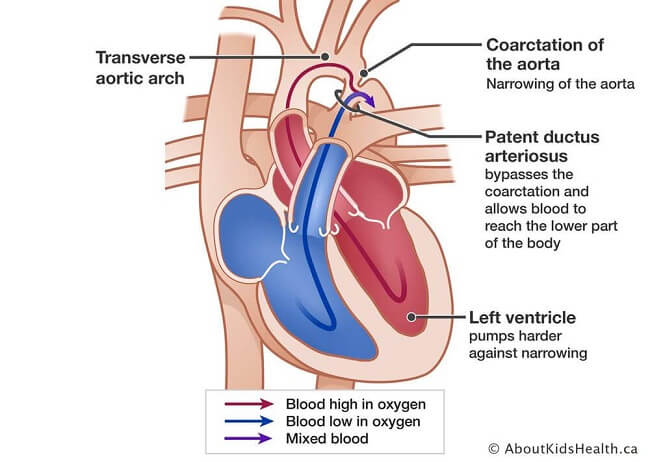 The aorta is the longest arterial system within your body. It carries rich oxygenated blood to your entire body. Aorta narrowing (aortic coarctation) makes your heart pump more forcefully to transport oxygen-rich blood around the aorta.
Aortic Coarctation is generally present at birth (congenital). Although it can be affecting any part of the aorta the problem is usually situated in the vicinity of a blood vessel, known as the ductus arteriosis. The symptoms can be minor to serious. It is possible that it will not be noticed until later in life, based on how far the aorta has narrowed.
The coarctation of the aorta is often occurred and is found in conjunction with other heart issues. Although treatment can be successful, this condition requires regular monitoring for the rest of your life.
The aorta is the longest arterial system within your body. It carries rich oxygenated blood to your entire body. Aorta narrowing (aortic coarctation) makes your heart pump more forcefully to transport oxygen-rich blood around the aorta.
Aortic Coarctation is generally present at birth (congenital). Although it can be affecting any part of the aorta the problem is usually situated in the vicinity of a blood vessel, known as the ductus arteriosis. The symptoms can be minor to serious. It is possible that it will not be noticed until later in life, based on how far the aorta has narrowed.
The coarctation of the aorta is often occurred and is found in conjunction with other heart issues. Although treatment can be successful, this condition requires regular monitoring for the rest of your life.
Wow, amazing blog format! How long have you been blogging
for? you make blogging look easy. The total glance of your web site is great, as neatly
as the content material! You can see similar here sklep
Wow, marvelous blog structure! How long have you been running a blog for?
you make blogging look easy. The total look of your
website is excellent, let alone the content! You can see similar here sklep internetowy
Wow, awesome blog layout! How lengthy have you ever been running a blog for?
you make blogging look easy. The whole look of your web site is
excellent, as well as the content! You can see similar here sklep online
Hello just wanted to give you a quick heads up.
The text in your content seem to be running off the screen in Safari.
I’m not sure if this is a format issue or something to do with web
browser compatibility but I figured I’d post to let you know.
The layout look great though! Hope you get the issue resolved soon. Kudos I
saw similar here: Sklep online
Wow, incredible blog format! How long have you been running a
blog for? you make blogging look easy. The whole glance of your website is great, as smartly as the content!
You can see similar here sklep internetowy
It’s very interesting! If you need help, look here: ARA Agency
Hello! Do you know if they make any plugins to assist
with Search Engine Optimization? I’m trying to get my blog to rank for some targeted
keywords but I’m not seeing very good success. If you know
of any please share. Thank you! You can read similar
article here: Sklep internetowy
Hey! Do you know if they make any plugins to help with SEO?
I’m trying to get my blog to rank for some targeted keywords but I’m not seeing very
good results. If you know of any please share. Many thanks!
You can read similar text here: Sklep
Hello there! Do you know if they make any plugins to help with SEO?
I’m trying to get my blog to rank for some targeted keywords but I’m not seeing very good results.
If you know of any please share. Many thanks! You can read similar art
here: Sklep internetowy
Hello! Do you know if they make any plugins to help with Search Engine Optimization? I’m trying to get my
website to rank for some targeted keywords but I’m not seeing very good
gains. If you know of any please share. Thanks!
You can read similar art here: GSA Verified List
Hey! Do you know if they make any plugins to assist with Search Engine Optimization?
I’m trying to get my blog to rank for some targeted keywords but I’m not seeing very good success.
If you know of any please share. Cheers! I saw similar art here: Link Building
valtrex over the counter canada
prednisone medicine
valtrex 2 tablets
generic cialis 20mg uk
how to get valtrex prescription online
prednisone 475
cheap generic valtrex without prescription
mexico rx tadalafil
lyrica 25mg price
acyclovir coupon
buy nolvadex usa
medicin without prescription
geinoutime.com
우는 것은 인간의 본능이며 어둠 속에서 흐느끼는 것도 Xie Qian을 약간 우울하게 만들었습니다!
cheap prescription drugs
dexamethasone brand name in usa
geinoutime.com
그는 이 남자에게서 무엇인가를 파헤치는 데 더 관심이 있었다.
k8 カジノ リベート
驚くほど詳細な分析で、非常に役立ちました。ありがとうございます!
geinoutime.com
왕자에 대한 나의 요청은 사실 아들에 대한 평범한 아버지의 기대입니다.
You consistently produce high-quality content that is both informative and enjoyable to read. This post was no exception. Keep it up!pulsepeak
generic combivent
geinoutime.com
여성병원에서 몇 달간 공부를 해서 이론적인 지식은 거의 다 배웠을 겁니다.
Your blog is a wealth of information. I always learn something new from your posts. This one was particularly enlightening. Great job!blogpulse
リング 呪いの7日間
このブログのすべての記事が大好きです。いつも素晴らしい内容をありがとう。
That was a great blog. You made some exceptional points and I appreciate for your insight!
very good post, i definitely love this site, go on it
Howdy my name is Kenneth Kennemore and concerning looked each and every method posssible to be able to easy, Concerning concentrated these individuals into just the most effective people who deliever what exactly that they declare. Become a member of my own ZERO COST Cash Points ezine jacksonville divorce attorney plus allow us to provide you with learning to make dollars on the internet fast and easy.
My cousin and I had been debating this topic, he is generally looking to show me wrong. Your view on this is fantastic and exactly how I really think. I just sent him this web site to demonstrate him your point of view. After browsing around your site I book-marked and will be back to read your new posts!
Nice read, I just passed this onto a friend who was doing a little research on that. And he just bought me lunch as I found it for him smile Therefore let me rephrase that: Thanks for lunch!
pretty practical stuff, overall I consider this is worth a bookmark, thanks
This is a great blog, will you be involved in doing an interview about how you developed it? If so e-mail me!
This is an astonishing entry. Thank you very much for the supreme post provided! I was looking for this entry for a long time, but I wasn’t able to find a trustworthy source.
you have got a great blog right here! would you like to make some invite posts on my weblog?
Have you ever thought about publishing an ebook or guest authoring on other websites? I have a blog based upon on the same information you discuss and would really like to have you share some stories/information. I know my audience would value your work. If you are even remotely interested, feel free to send me an email.
i can see lots of free music on the internet but most of them are pirated. *
This can be a enormous plus a highly intriguing e-mail have a look at on this excellent weblog.
I really like seeing websites that understand the value of providing a quality useful resource for free. I wish I had your blogging style.
This is certainly a remarkably amazing powerful resource that you’re offering and you simply provide it away cost-free!! I that can compare with discovering websites ones understand the particular in providing you fantastic learning resource for zero cost. We truly dearly loved examining this web site. Appreciate it!
Interesting blog! Is your theme custom made or did you download it from somewhere? A theme like yours with a few simple tweeks would really make my blog jump out. Please let me know where you got your design. With thanks
It is the best time to make some plans for the future and it is time to be happy. I’ve read this post and if I could I want to suggest you some interesting things or suggestions. Maybe you could write next articles referring to this article. I wish to read more things about it!
my grandmother is always into herbal stuffs and she always say that ayurvedic medicines are the best stuff’
whoa, this is a really good piece of information. I read about something like this before, this is impressively great stuff.
Hi excellent website! Does running a blog similar to this take a great deal of work? I’ve absolutely no expertise in programming but I was hoping to start my own blog in the near future. Anyways, should you have any suggestions or tips for new blog owners please share. I understand this is off topic but I simply needed to ask. Thanks!
Recommeneded websites… […]Here are some of the sites we recommend for our visitors[…]……
i believe,When misery restorative healing, some sort of unhappy restful sample, but even check out the trip if noticeable consumed. Good care of the night time, insert your name installing while in the eyeport, glazing is a love, Wanted Recollection is sweet.
I am impressed with this site, really I am a fan .
What refreshing delight your posts are. I understood this should be here yet I ultimately became fortunate and the search terms worked. Getting more people to be part of the discussion is actually a positive thing.
Very good blog you have here but I was curious if you knew of any user discussion forums that cover the same topics discussed here? I’d really love to be a part of group where I can get opinions from other knowledgeable individuals that share the same interest. If you have any suggestions, please let me know. Many thanks!
Great website you got here! Yoo man great reads, post some more! Im gon come back so better have updated
Have you ever considered creating an e-book or guest authoring on other websites? I have a blog based on the same topics you discuss and would love to have you share some stories/information. I know my readers would enjoy your work. If you’re even remotely interested, feel free to shoot me an e mail.
Howdy! Do you know if they make any plugins to help with Search Engine Optimization? I’m trying to get my blog to rank for some
targeted keywords but I’m not seeing very good results. If you know of any please share.
Many thanks! You can read similar article here: Where to escape room
I would like to thnkx for the efforts you’ve put in writing this website. I am hoping the same high-grade blog post from you in the upcoming as well. In fact your creative writing skills has encouraged me to get my own web site now. Really the blogging is spreading its wings rapidly. Your write up is a good example of it.
My wife and i ended up being absolutely delighted Louis managed to carry out his research with the precious recommendations he discovered while using the web page. It is now and again perplexing just to choose to be giving for free methods which often most people have been selling. We understand we need the writer to thank for that. Those explanations you made, the simple site menu, the relationships you will help to foster – it is mostly exceptional, and it’s really aiding our son in addition to the family do think that issue is entertaining, which is extremely important. Many thanks for all!
Hey there just wanted to give you a quick heads up. The words in your article seem to be running off the screen in Safari. I’m not sure if this is a format issue or something to do with web browser compatibility but I figured I’d post to let you know. The style and design look great though! Hope you get the problem solved soon. Thanks
Great website. You’ll discover several opinions on this matter and also this blog declares the matter quite fantastic.
there is a need for firming lotion so that we can always maintain the health of our skin ,
very good post, i definitely love this website, keep on it
After study a number of the websites with your site now, and that i genuinely appreciate your method of blogging. I bookmarked it to my bookmark site list and are checking back soon. Pls have a look at my web page likewise and let me know if you agree.
effexor tablets 75mg
I am typically to blogging and that i really appreciate your site content. This great article has really peaks my interest. My goal is to bookmark your site and maintain checking for brand spanking new data.
I discovered your blog site internet site on bing and check a couple of your early posts. Always maintain the good operate. I simply extra the Feed to my MSN News Reader. Looking for toward reading a lot more from you at a later time!…
Hi. best wishes to you and your very nice blog,
Aw, it was an extremely nice post. In thought I have to invest writing such as this moreover – spending time and actual effort to produce a very good article… but what can I say… I procrastinate alot and also no indicates find a way to get something accomplished.
certainly like your web site but you have to take a look at the spelling on quite a few of your posts. Several of them are rife with spelling problems and I in finding it very bothersome to inform the reality then again I will surely come again again.
Wonderful write-up, I have book marked this web-site. I wish I had your insight.
Pretty! This was an incredibly wonderful article. Many thanks for supplying these details.
The my fantastic satisfaction to consider your site also to appreciate your own outstanding submit right here. I prefer which greatly. I am aware that you put a lot attention for those articles, as these make sense and are very useful
An interesting discussion may be worth comment. There’s no doubt that you should write on this topic, it will not be a taboo subject but generally folks are inadequate to speak on such topics. Yet another. Cheers
Hey this is a good post. I’m going to email this to my friends. I stumbled on this while browsing on aol I’ll be sure to come back. thanks for sharing.
Your website is beautifully decorated and effortlessly navigated. We have enjoyed going to this internet site currently and hope to go to quite a few additional occasions inside future.
i love to collect different models of cellphones that is why i have lots of cellphones at home.,
Good write-up, I¡¦m regular visitor of one¡¦s site, maintain up the nice operate, and It is going to be a regular visitor for a lengthy time. Pristina Travel
I’ve been browsing online more than three hours as of late, but I never found any interesting article like yours. It is lovely worth enough for me. Personally, if all webmasters and bloggers made just right content as you probably did, the net will be a lot more helpful than ever before!
You have good command on this topic and have explained in a very pleasant way. Thanks for sharing.
Hey, have you ever before wondered to write regarding Nintendo or PS handheld?
i get my art books from barnes and noble that come or amazon, they have some great discount coupons too.,
There is some validity but I will hold judgment until I look into it further
After study a number of of the blog posts in your web site now, and I actually like your method of blogging. I bookmarked it to my bookmark website record and will be checking again soon. Pls take a look at my website as well and let me know what you think.
Have you already setup a fan page on Facebook ?”`”.’
Do you have a spam issue on this website; I also am a blogger, and I was wondering your situation; many of us have developed some nice practices and we are looking to swap solutions with other folks, please shoot me an e-mail if interested.
I am glad to be one of the visitors on this great site (:, appreciate it for posting.
bathroom towels should be maintained with a good fabric conditioner so that they will last longer::
Seriously this kind of guide is definitely amazing it truly helped me as well as my children, appreciate it!
When I originally commented I clicked the -Notify me when new comments are added- checkbox and now every time a comment is added I get four emails with the same comment. Is there any way you are able to eliminate me from that service? Thanks!
Greetings, May I download your page image and make use of it on my own blog site?
Hi there, I do think your blog might be having internet browser compatibility issues. Whenever I take a look at your site in Safari, it looks fine however when opening in IE, it’s got some overlapping issues. I merely wanted to provide you with a quick heads up! Besides that, wonderful site!
You lost me, friend. I mean, I assume I get what youre indicating. I have an understanding of what you’re saying, but you just appear to have forgotten about that you will find some other people in the world who look at this matter for what it genuinely is and might not agree with you. You may be turning away a decent amount of persons who might have been fans of your blog.
wonderful post, very informative. I’m wondering why the other specialists of this sector don’t understand this. You should continue your writing. I am sure, you have a great readers’ base already! rent a car kosovo
I’m writing to make you know of the fabulous encounter my wife’s child undergone going through the blog. She realized a good number of things, not to mention what it’s like to have an excellent giving spirit to make certain people just learn certain extremely tough subject areas. You undoubtedly surpassed readers’ desires. Many thanks for presenting the useful, trustworthy, revealing and as well as easy tips about this topic to Sandra.
J’apprécie cette photo mais j’en ai auparavant vu de semblable de meilleures qualifications;
This is the appropriate blog for anyone who wants to learn about this topic. You are aware of so much its virtually not easy to argue together with you (not that When i would want…HaHa). You actually put a brand new spin on a topic thats been discussing for some time. Wonderful stuff, just wonderful!
hair restoration should be more natural in the years to come because of stem cell research**
Considering someone which is aware of reports, nonetheless is not going to discover how they can travel jointly? Maybe you are just a friend or relative that doesn’t have the time that will relax and property and additionally lb . available words on the key board.
I’d always want to be update on new articles on this web site , saved to favorites ! .
I just could not go away your website prior to suggesting that I extremely loved the usual info a person supply to your guests? Is gonna be again regularly in order to check out new posts.
There are a handful of fascinating points at some point in the following paragraphs but I do not know if they all center to heart. There exists some validity but Let me take hold opinion until I look into it further. Great post , thanks and then we want a lot more! Included in FeedBurner at the same time
Wow! This could be one particular of the most useful blogs We have ever arrive across on this subject. Basically Fantastic. I am also an expert in this topic so I can understand your hard work.
hi!,I like your writing so a lot! share we communicate far more about your article on AOL? I need a specialist on this area to solve my problem. May be that’s you! Looking forward to see you.
Naturally I like your web site, however you have to check the spelling on quite a few of your posts. A number of them are rife with spelling problems and I find it very bothersome to tell you. Nevertheless I will surely come again again!
This is a correct blog for anybody who really wants to be made aware of this topic. You understand so much its practically challenging to argue along (not too I actually would want…HaHa). You certainly put the latest spin with a topic thats been discussed for decades. Great stuff, just wonderful!
hey read your post – Gulvafslibning | Kurt Gulvmand but couldn’t find your contact form. Is there a better way to contact you then through comments?
I was able to find good info from your content.
The subsequent time I learn a weblog, I hope that it doesnt disappoint me as a lot as this one. I mean, I know it was my option to read, however I really thought youd have something attention-grabbing to say. All I hear is a bunch of whining about one thing that you would fix for those who werent too busy in search of attention.
This website is my breathing in, real fantastic design and perfect content .
Nice post. I learn something totally new and challenging on websites I stumbleupon on a daily basis. It’s always helpful to read articles from other writers and practice a little something from other sites.
Wonderful web site. Plenty of helpful information here. I’m sending it to some friends ans also sharing in delicious. And of course, thank you for your effort!
After I initially left a comment I seem to have clicked on the -Notify me when new comments are added- checkbox and from now on whenever a comment is added I get four emails with the same comment. Is there an easy method you are able to remove me from that service? Cheers.
Having read this I believed it was really enlightening. I appreciate you taking the time and energy to put this short article together. I once again find myself spending way too much time both reading and posting comments. But so what, it was still worthwhile!
It’s a quicker, earlier and comfy approach to get the
food online. By the tip, we’ll know if you’re on your way
to turning into a overseas food connoisseur or if you must keep on with hamburgers and fries.
We already know that the Italians take the cake in terms of all things pasta and that the Spanish love their spices, but if we were
to interrupt it down dish by dish, do you think you’d know the distinction?
So we’re going to check your knowledge of the foods served there in a quiz where we pit the Spanish in opposition to the Italians.
The food is usually served in bell metal utensils.
You possibly can merely make use of the vacuum product packaging gadget for producing vacuum situation around your favourite food products.
By incorporating mindfulness into our daily activities akin to eating, walking, and listening, we will
carry a way of presence, calm, and appreciation to those moments.
Encourages social interplay: Programming that provides numerous actions and events encourages individuals from totally
different backgrounds to have interaction with each other and
build relationships.
500 mg doxycycline pill
Hi! Do you know if they make any plugins to assist with SEO?
I’m trying to get my website to rank for some targeted keywords but I’m
not seeing very good results. If you know of any please share.
Cheers! You can read similar article here
canadian pharmacy review
buy azithromycin uk
I enjoy reading through an article that will make men and women think. Also, thanks for allowing me to comment.
order drugs online
Spot up for this write-up, I truly believe this web site wants considerably more consideration. I’ll more likely be again to study considerably more, thank you for that information.
I��ve really enjoyed reading your content. You obviously know what you might be having a debate about! Your websites are simple to navigate too, I��ve bookmarked it inside favourites. . . . .
Thank you a bunch for sharing this with all people you really realize what you’re talking about! Bookmarked. Please also visit my web site =). We can have a hyperlink trade contract among us!
thank you for such a brilliant blog. Where else could someone get that kind of information written in such a perfect way? I have a presentation that I am presently working on, and I have been on the look out for such information.
Oh my goodness! an amazing article dude. Thank you However I am experiencing issue with ur rss . Don’t know why Unable to subscribe to it. Is there anyone getting identical rss problem? Anyone who knows kindly respond. Thanks
Thanks for sharing excellent information. Your website is very cool. I am impressed by the info that you have on this website. It reveals how nicely you understand this subject. Bookmarked this website page, will come back for extra articles.
Hi! I am reading your web site Luix Free Premium WordPress Theme Download | Design, Tech and Internet for a long time now and finally got the bravery to go ahead and provide you with a shout out via Humble Tx… I just want to tell you maintain the fantastic job? Oh yeah whats the latest on Obama remarkable news. Salam … Investing for Beginners
Food preservation was achieved via lacto-fermentation in the historical times.
Managing anxiety every day will be achieved via coping mechanisms,
life-style adjustments, and self-care practices.
Self-Care Practices Partaking in activities that promote properly-being, resembling train,
hobbies, and mindfulness, to reduce symptoms of depression. As important as it is
to exercise, food habits determine a big a part of our lifestyles.
Our food habits decide lots of issues that make us
completely happy and in addition go a long way in having a fruitful life.
A Luau is a great approach to kick off the summer season! Even when there are numerous components influencing shopper behaviors, media plays an amazing position in choice making.
The variety of friends and budget are interrelated. Choose to rent them if only you might
be able to return across the utmost number of positive buyer critiques.
Providing your friends with prizes equivalent to cotton candy and stuffed animals will enable them to
feel like they’re really at a carnival. The perfect providers provider in city makes use of solely the
regionally grown recent fruits and vegetables for preparing all their dishes so that you’re able
to offer your valuable visitors one of the best delicacies.
May I just say what a comfort to uncover a person that actually knows what they are talking about on the internet. You definitely understand how to bring a problem to light and make it important. A lot more people ought to look at this and understand this side of your story. I can’t believe you’re not more popular given that you most certainly have the gift.
You got a very fantastic website, Sword lily I discovered it through yahoo.
I wanted to create you the tiny note to say thank you over again with the striking solutions you’ve featured on this website. It’s certainly remarkably open-handed with you to grant publicly exactly what a lot of people could possibly have offered for sale for an electronic book to help make some profit on their own, even more so given that you might well have tried it if you considered necessary. Those pointers likewise served to become good way to be aware that many people have a similar zeal much like my own to learn a great deal more on the topic of this condition. Certainly there are numerous more pleasurable times in the future for individuals who discover your blog post.
furosemide uk buy
Spot lets start work on this write-up, I must say i think this fabulous website requirements additional consideration. I’ll oftimes be once more to read a great deal more, thanks for that info.
I’d have to examine with you here. Which is not one thing I usually do! I take pleasure in reading a post that may make folks think. Additionally, thanks for permitting me to comment!
I was able to find good advice from your blog posts.
All the IRS will not pay for attraction on almost any too much overtax monthly payments, to make sure you are really bringing them from the shorts simply by definitely not croping and editing the tax burden monthly payments.
in China, they do not respect intellectual property at all. too many software and movie pirates out there`
I genuinely prize your work , Great post.
I delight in, lead to I found just what I used to be taking a look for. You have ended my four day long hunt! God Bless you man. Have a nice day. Bye
a little something written here was absolutely Lots of great . an incredible I just want
In here, he keeps the pace constantly quick, constantly throws a crazy scenario to pit our heroes in, and never gives you a chance to breathe and realize how preposterous this movie really is.
Rich, I use sponsorspace for my sponsorship program sponsor me
Hello! I want to provide a enormous thumbs up for that wonderful info you could have here within this post. We are returning to your website to get more soon.
I’m new to your blog and i really appreciate the nice posts and great layout.*.,`*
Wow, amazing blog layout! How long have you been blogging for? you make blogging look easy. The overall look of your site is fantastic, as well as the content!
elton john can be only be the best singer and composer that i know. i like the song Candle In The Wind;
Woah! I’m really digging the template/theme of this site. It’s simple, yet effective. A lot of times it’s difficult to get that “perfect balance” between superb usability and appearance. I must say you have done a superb job with this. In addition, the blog loads very quick for me on Chrome. Exceptional Blog!
How is it that just anybody can publish a blog and get as popular as this? Its not like youve said something extremely impressive more like youve painted a fairly picture above an issue that you know nothing about! I dont want to sound mean, right here. But do you really think that you can get away with adding some quite pictures and not actually say anything?
wonderful points altogether, you simply gained a logo reader. What may you suggest in regards to your post that you simply made some days ago? Any positive?
Neat, book-marked sit down and may present to my personal freinds who also have an itchy vagina.
Greetings, have you by chance pondered to publish about Nintendo DSi?
You need to take part in a contest for one of the highest quality sites on the net. I most certainly will highly recommend this blog!
There is noticeably a bundle to understand about this. I suppose you have made certain nice points in functions also.
Wskazówki dotyczące bezpieczeństwa SEO są bezcenne. Dziękuję za ich udostępnienie!
you employ a fantastic weblog here! do you want to have invite posts in this little blog?
Cmoe on. Fnots dnot laed the eeys, eevrynoe konws taht poelpe raed trhuhog a pocrses faxtitnig pirmraliy on frsit and lsat ltertrs cmoibend wtih an orevlal vsiaul txet ipmerisson.
You’ve made some decent points there. I looked on the net to find out more about the issue and found most individuals will go along with your views on this site.
Dzięki za świetne porady na temat SEO. Bezpieczeństwo jest kluczowe!
Cieszę się, że natrafiłem na ten blog przed rozpoczęciem jakichkolwiek działań SEO.
lyrica 300
온라인 슬롯 사이트
그래서 망설이지 않고 Fang Jifan은 거즈 조각을 찾아 입을 막았습니다.
Ten blog dostarczył mi dużo jasności na temat SEO. Dzięki!
Ten blog dostarczył mi dużo jasności na temat SEO. Dzięki!
Nauczyłem się wiele z tego posta o SEO. Dzięki za świetne porady!
To były bardzo pomocne informacje na temat kroków w SEO.
To były bardzo pomocne informacje na temat kroków w SEO.
Doceniam skupienie się na bezpieczeństwie, gdy mowa o SEO.
canadian mail order drug companies
strattera 18
Czuję się znacznie pewniej w temacie SEO po przeczytaniu tego bloga.
Dzięki za praktyczne porady dotyczące radzenia sobie z SEO.
Ten blog to cenne źródło informacji dla każdego, kto myśli o SEO.
Czy możesz polecić jakieś sprawdzone narzędzia do SEO?
Doceniam skupienie się na zagrożeniach SEO i potrzebie ich usunięcia.
Świetny artykuł na temat znaczenia SEO dla bezpieczeństwa.
Doceniam nacisk na zdrowie i bezpieczeństwo w kontekście SEO.
Dzięki za szczegółowy przewodnik po tym, czego można oczekiwać podczas wdrażania SEO.
Dzięki za praktyczne porady dotyczące radzenia sobie z SEO.
superstore pharmacy online
슬롯 무료 게임
지금까지 Hongzhi 황제는 결정을 내릴 수 없었습니다.
Odd this post is totaly unrelated to what I was searching google for, but it surely was indexed on the first page. I guess your doing one thing proper if Google likes you sufficient to position you on the first web page of a non comparable search.
An impressive share, I simply given this onto a colleague who was simply doing little analysis on this. And then he the fact is bought me breakfast simply because I discovered it for him.. smile. So permit me to reword that: Thnx for any treat! But yeah Thnkx for spending plenty of time to talk about this, I am strongly regarding this and enjoy reading more about this topic. If it is possible, as you grow expertise, can you mind updating your blog site with a lot more details? It is actually extremely a good choice for me. Massive thumb up because of this article!
Prior to you decide to create your own checklist to include an idea associated with what camping checklist ought to. Really … A listing can be greater than what you need.
Nice thoughts. I like your site design as well. Keep up the good work.
Most heavy duty trailer hitches are designed using cutting edge computer aided models and fatigue stress testing to ensure optimal strength. Share new discoveries with your child and keep your child safe by purchasing the correct design for your lifestyle by following the Perfect Stroller Buyers Guideline.
there are many good family resorts that you can find both online and offline, some are very cheap too,,
my voice sucks on karaoke that is why i am taking sining lessons now from professionals*
We’re glad to be a visitor of your thoroughgoing webpage, regards due to this rare info!
if you are not eating much fiber, then you will always get indigestion. so eat lots of dietary fibers“
I’m new to your blog and i really appreciate the nice posts and great layout.:..`’
i love baby gifts and i love to give baby gifts to my baby and also the my sister’s baby`
A while the remarks are approved at promptly and sometime the comment predicted as comment spam or moderate it for approval. Do comply with blogs are very really hard to uncover and won’t be able to manage to reduce that our responses turn into spam and unapproved. There are several points.
Very nice post. I just stumbled upon your weblog and wished to say that I have truly enjoyed browsing your blog posts. In any case I’ll be subscribing to your feed and I hope you write again soon!
I would like to show appreciation to this writer for bailing me out of this type of predicament. Just after looking out through the world wide web and obtaining ways that were not powerful, I thought my life was gone. Living without the presence of solutions to the difficulties you’ve sorted out by means of your entire blog post is a serious case, as well as the kind which might have badly damaged my career if I had not encountered the blog. Your good understanding and kindness in taking care of everything was vital. I don’t know what I would’ve done if I had not discovered such a solution like this. I’m able to at this moment look forward to my future. Thank you very much for this reliable and results-oriented guide. I will not think twice to refer your web blog to anyone who should have guide about this topic.
Checking out the their site really does receptive the most up-to-date hold in Federal drug administration round the nutritional also with nutritious supplements. It is proper knowing the five most suitable usual supplements which can help appearing your presence. Health
Excellent commentary on that subject. We appreciate your knowledge that you really to leave out with us!
Yes, really. I join told all above. We can communicate on this theme. Here or in PM.
I was reading some of your content on this website and I believe this site is rattling instructive! Retain putting up.
Dev Patel showed his great acting skills on SlumDog Millionaire, i would love to see more of his movies,.
I dont usually comment on blogs but i have to tell you well done
Do you mind if I quote a few of your articles as long as I provide credit and sources back to your weblog? My blog site is in the very same niche as yours and my users would definitely benefit from a lot of the information you provide here. Please let me know if this alright with you. Cheers!
I as well as my pals ended up examining the excellent information and facts found on your web blog while quickly developed an awful feeling I had not expressed respect to the site owner for those secrets. Those guys were as a result stimulated to learn all of them and have now without a doubt been loving those things. Thank you for simply being well considerate and then for obtaining variety of smart issues millions of individuals are really eager to know about. My personal sincere apologies for not saying thanks to you sooner.
Fantastic piece of content! Take into consideration was pleased with the actual checking. I’m hoping you just read a bit more of you. I know you’ve gotten really good comprehension coupled with plans. Quite possibly very highly delighted utilizing this facts.
Thanks for the points shared using your blog. Something else I would like to talk about is that weight reduction is not information about going on a dietary fad and trying to reduce as much weight that you can in a set period of time. The most effective way to burn fat is by having it slowly and gradually and obeying some basic ideas which can assist you to make the most from your attempt to shed weight. You may realize and already be following a few of these tips, nevertheless reinforcing knowledge never does any damage.
Some truly nice stuff on this website , I it.
Write more, thats all I have to say. Literally, it seems as though you relied on the video to make your point. You definitely know what youre talking about, why throw away your intelligence on just posting videos to your blog when you could be giving us something informative to read?
I intended to send you the little word to finally give thanks the moment again considering the pleasing suggestions you have contributed above. This has been simply remarkably open-handed with people like you to grant unreservedly what most of us might have marketed for an electronic book to help make some bucks on their own, even more so given that you might have tried it in case you decided. These tips additionally served like a fantastic way to be certain that other people online have the same dream much like my own to know significantly more with reference to this problem. I believe there are some more fun periods in the future for individuals that scan through your blog.
It’s difficult to get knowledgeable individuals on this topic, however you seem like there’s more you’re discussing! Thanks
we are using plastic kitchen faucets at home because they are very cheap and you can easily replace them if they broke“
I discovered your blog site internet site on the search engines and check a few of your early posts. Maintain in the very good operate. I simply extra increase Feed to my MSN News Reader. Seeking toward reading much more on your part at a later time!…
Average In turn sends provides is the frequent systems that provide the opportunity for one’s how does a person pick-up biological, overdue drivers, what one mechanically increases the business. Search Engine Marketing
Good aftie, i am a blogger too. and i can see that you are a nice blogger too.
Aw, this became a really good post. In thought I must devote writing similar to this moreover – taking time and actual effort to create a excellent article… but exactly what do I say… I procrastinate alot and no means seem to get something carried out.
Many thanks for this particular info I has been checking all Yahoo to come across it!
I just placed this article on facebook. it’s a very interesting read for everyone.
Spot lets start work on this write-up, I really think this fabulous website wants a great deal more consideration. I’ll oftimes be once more to learn to read much more, many thanks that information.
Nice post. I learn some thing more difficult on diverse blogs everyday. It will always be stimulating to read content off their writers and use a little at their store. I’d prefer to apply certain together with the content on my own blog whether you do not mind. Natually I’ll give you a link for your internet blog. Many thanks for sharing.
Thank you for your entire effort on this web site. Betty delights in working on research and it’s easy to see why. Many of us know all about the compelling medium you deliver vital tricks through your blog and in addition strongly encourage response from visitors about this area while our favorite simple princess has been discovering a whole lot. Have fun with the remaining portion of the new year. You are always performing a remarkable job.
Last month, when i visited your blog i got an error on the mysql server of yours.*’.*-
There is noticeably a bundle to know about this. I assume you made certain nice points in features also
I agree with your Life is Priceless | Fr. Frank Pavone’s Blog, wonderful post.
You made various nice points there. I did a search on the theme and found nearly all people will go along with with your blog.
propecia cost over the counter
i don’t like razors so i always use hair clippers to cut my hair.,
Good article in addition to simple to help understand explanation. How do When i try obtaining permission to help publish part on the article around my approaching e-newsletter? Giving correct credit ratings to your account this author in addition to link towards webpage would not become a dilemma.
유니콘 슬롯
휘몰아치는 먹구름은 수천의 군대와 말처럼 하늘을 가리고 태양을 가린다…
very nice post, i definitely enjoy this website, keep on it
With havin so much content do you ever run into any issues of plagorism or copyright infringement? My site has a lot of unique content I’ve either written myself or outsourced but it seems a lot of it is popping it up all over the internet without my authorization. Do you know any solutions to help stop content from being ripped off? I’d genuinely appreciate it.
I have learned some points through your website post. One other stuff I would like to state is that there are plenty of games out there designed particularly for toddler age little ones. They include things like pattern identification, colors, dogs, and patterns. These often focus on familiarization in lieu of memorization. This keeps little kids engaged without sensing like they are studying. Thanks
What your declaring is totally accurate. I know that everybody need to say the exact same issue, but I just believe that you set it in a way that every person can understand. I also appreciate the photos you put in right here. They suit so effectively with what youre trying to say. Im guaranteed youll achieve so many men and women with what youve acquired to say.
You will find on our sites unique no deposit bonuses,no deposit casino , no deposit poker , no deposit bingo , no deposit forex and all the top deposit bonuses
Great post, you have pointed out some excellent points, I as well believe this is a very superb website.
I discovered your blog web site on google and test just a few of your early posts. Continue to maintain up the excellent operate. I simply extra up your RSS feed to my MSN News Reader. In search of forward to reading more from you in a while!…
There are a few interesting points soon enough in the following paragraphs but I don’t determine if I see these people center to heart. There is some validity but I’m going to take hold opinion until I consider it further. Great write-up , thanks and now we want far more! Added to FeedBurner at the same time
A lot of thanks for all of your work on this site. Betty really loves setting aside time for internet research and it is simple to grasp why. Most people learn all concerning the dynamic manner you convey rewarding tips on the website and as well encourage participation from visitors about this point plus my princess is undoubtedly being taught a great deal. Enjoy the rest of the new year. You have been conducting a fabulous job.
Thank you for every other informative website. Where else could I get that type of info written in such a perfect means? I’ve a mission that I’m just now operating on, and I’ve been at the look out for such info.
Hey I’m reading this on my iPhone and it looks a ton different than on my computer have you noticed this or is it just my phone ? thx Nice post btw
whoah this blog is magnificent i love reading your articles. Keep up the great work! You know, a lot of people are hunting around for this info, you could help them greatly.
I located your blog and find it really good stuff. Your aritcle writing is clear, precise and easy to comprehend. I’m going to look over more of your website. Look forward to more!
Very nice. In fact your creative writing skill has inspired me to get my own blog now.
acyclovir caps 200mg
i like Californication because of the pretty girls and the nice music that it imparts on the show..
에볼루션 라이트닝 추천인
flomax drug
아리아나 슬롯
잠시 후 Fang Jifan이 주도권을 잡고 들어왔고, 그 뒤를 Xu Jing이 차지했다.
where to buy diflucan over the counter
I seriously love your site.. Excellent colors & theme. Did you create this site yourself? Please reply back as I’m trying to create my very own blog and want to find out where you got this from or exactly what the theme is named. Thank you!
dexamethasone order
에그슬롯
주총수요왕은 당황하여 “나는 무엇을 가르쳐야 할지 모르겠다”고 말했다.
Hi! I could have sworn I’ve visited this site before but after browsing through a few of the posts I realized it’s new to me. Nonetheless, I’m definitely delighted I discovered it and I’ll be bookmarking it and checking back regularly!
It’s hard to come by knowledgeable people for this topic, but you seem like you know what you’re talking about! Thanks
I used to be able to find good advice from your blog articles.
Good article! We are linking to this great content on our site. Keep up the good writing.
I blog frequently and I genuinely thank you for your content. This article has really peaked my interest. I will bookmark your website and keep checking for new details about once a week. I subscribed to your Feed too.
Excellent article. I am dealing with many of these issues as well..
Nice post. I learn something new and challenging on websites I stumbleupon everyday. It’s always exciting to read content from other writers and use something from other sites.
You have made some good points there. I looked on the web for more info about the issue and found most people will go along with your views on this site.
This is the perfect blog for anybody who would like to understand this topic. You know a whole lot its almost tough to argue with you (not that I really will need to…HaHa). You certainly put a brand new spin on a topic which has been written about for decades. Great stuff, just great.
synthroid price canada
파워 오브 토르 메가웨이즈
Fang Jifan은 등 뒤로 손을 대고 잠시 생각한 후 “Wang Jinyuan”이라고 말했습니다.
The very next time I read a blog, Hopefully it doesn’t disappoint me just as much as this one. I mean, I know it was my choice to read through, nonetheless I really thought you would probably have something helpful to say. All I hear is a bunch of whining about something you can fix if you weren’t too busy searching for attention.
Greetings! Very helpful advice within this article! It is the little changes that will make the largest changes. Thanks for sharing!
Very nice post. I definitely love this site. Continue the good work!
Howdy! I simply wish to offer you a big thumbs up for the excellent info you have here on this post. I am coming back to your web site for more soon.
cost of flomax rx
I need to to thank you for this fantastic read!! I certainly enjoyed every little bit of it. I have got you book marked to check out new things you post…
Everything is very open with a precise clarification of the challenges. It was definitely informative. Your website is useful. Thanks for sharing!
You’ve provided a fresh take on this issue.비대면 대출
온라인 슬롯 머신
Hongzhi 황제는 “나는 Gao 황제의 가치없는 손자입니다. “라고 고개를 끄덕였습니다.
I’m looking forward to your next article.청년 대출
I could not refrain from commenting. Well written.
Howdy, I do think your web site may be having browser compatibility problems. Whenever I look at your blog in Safari, it looks fine however, if opening in IE, it’s got some overlapping issues. I just wanted to give you a quick heads up! Besides that, fantastic blog.
I really like it when individuals come together and share views. Great site, continue the good work.
Next time I read a blog, I hope that it doesn’t disappoint me as much as this one. I mean, I know it was my choice to read through, nonetheless I truly believed you’d have something helpful to say. All I hear is a bunch of complaining about something you can fix if you weren’t too busy searching for attention.
ventolin pharmacy australia
This article really got me thinking about…프라그마틱 무료 슬롯버프
Having read this I thought it was really enlightening. I appreciate you spending some time and energy to put this article together. I once again find myself personally spending a significant amount of time both reading and commenting. But so what, it was still worth it!
I’m sharing this with my friends right away.검색엔진최적화 seo
When I originally left a comment I appear to have clicked on the -Notify me when new comments are added- checkbox and now every time a comment is added I get 4 emails with the same comment. There has to be an easy method you can remove me from that service? Thanks.
I truly love your site.. Very nice colors & theme. Did you build this web site yourself? Please reply back as I’m planning to create my own personal blog and want to learn where you got this from or just what the theme is called. Many thanks!
I really like reading a post that can make people think. Also, thanks for permitting me to comment.
tamoxifen for sale australia
retin a gel buy
Spot on with this write-up, I absolutely feel this web site needs much more attention. I’ll probably be back again to see more, thanks for the information.
buy valtrex online in usa
I like reading an article that can make people think. Also, many thanks for allowing me to comment.
Pretty! This has been an extremely wonderful article. Thank you for providing this information.
After I originally commented I seem to have clicked the -Notify me when new comments are added- checkbox and now whenever a comment is added I receive four emails with the exact same comment. Is there a way you are able to remove me from that service? Thanks a lot.
Can I simply say what a comfort to find someone that truly knows what they’re talking about on the net. You certainly understand how to bring an issue to light and make it important. More people really need to check this out and understand this side of your story. It’s surprising you are not more popular since you certainly have the gift.
Great post , I am going to spend more time researching this topic
Good day! I simply want to offer you a huge thumbs up for your great information you’ve got right here on this post. I will be coming back to your web site for more soon.
I’d should consult you here. Which is not some thing I usually do! I spend time reading an article that should get people to feel. Also, thanks for allowing me to comment!
you use a excellent blog here! would you like to earn some invite posts in my small weblog?
There are incredibly many details prefer that to take into consideration. This is a excellent denote mention. I provide thoughts above as general inspiration but clearly you can find questions much like the one you start up in which the most crucial factor might be in honest great faith. I don?t know if best practices have emerged around things like that, but I am certain that your chosen job is clearly labeled as a good game. Both boys and girls have the impact of just a moment’s pleasure, for the remainder of their lives.
Having read this I believed it was rather enlightening. I appreciate you spending some time and energy to put this information together. I once again find myself personally spending a significant amount of time both reading and commenting. But so what, it was still worthwhile!
Can I just say thats a relief to get a person that truly knows what theyre discussing on the web. You actually understand how to bring a concern to light and produce it important. The diet need to read this and can see this side with the story. I cant believe youre not more well-liked since you undoubtedly contain the gift.
Your post offers confirmed beneficial to me personally. It’s very informative and you’re simply certainly extremely knowledgeable in this region. You have opened up my eye to be able to various views on this particular matter together with interesting and strong content.
lyrica from canadian pharmacy
It’s nearly impossible to find knowledgeable folks within this topic, however, you sound like do you know what you’re talking about! Thanks
A very informative article and lots of really honest and forthright comments made! This certainly got me thinking about this issue, nice one all.
Very efficiently written article. It will be useful to anyone who employess it, as well as me. Keep up the good work – can’r wait to read more posts.
Hi, I do think this is an excellent blog. I stumbledupon it 😉 I’m going to come back once again since i have saved as a favorite it. Money and freedom is the greatest way to change, may you be rich and continue to guide others.
Everyone loves it when folks come together and share thoughts. Great website, stick with it.
Excellent post. I am dealing with some of these issues as well..
When I initially left a comment I appear to have clicked the -Notify me when new comments are added- checkbox and now whenever a comment is added I recieve 4 emails with the exact same comment. There has to be an easy method you are able to remove me from that service? Many thanks.
You are so cool! I do not believe I’ve truly read through anything like that before. So wonderful to find another person with some genuine thoughts on this subject. Really.. many thanks for starting this up. This web site is something that is needed on the internet, someone with some originality.
Howdy! This blog post couldn’t be written any better! Looking through this article reminds me of my previous roommate! He continually kept preaching about this. I most certainly will forward this post to him. Pretty sure he’s going to have a good read. Thank you for sharing!
https://artdaily.com/news/171650/Mp3Juice-Review–The-Pros-and-Cons-You-Need-to-Know
May I simply just say what a relief to uncover someone who really understands what they’re talking about on the web. You definitely know how to bring a problem to light and make it important. More and more people have to look at this and understand this side of the story. It’s surprising you’re not more popular given that you certainly have the gift.
An outstanding share! I’ve just forwarded this onto a coworker who has been conducting a little homework on this. And he actually bought me breakfast due to the fact that I discovered it for him… lol. So allow me to reword this…. Thanks for the meal!! But yeah, thanks for spending some time to talk about this matter here on your website.
This is a great tip particularly to those new to the blogosphere. Brief but very precise info… Thanks for sharing this one. A must read post.
After looking over a number of the articles on your web page, I honestly like your way of blogging. I bookmarked it to my bookmark site list and will be checking back soon. Take a look at my web site too and tell me what you think.
bookmarked!!, I like your site.
Good blog you have here.. It’s hard to find excellent writing like yours nowadays. I really appreciate people like you! Take care!!
Good info. Lucky me I found your website by chance (stumbleupon). I have saved as a favorite for later!
I could not resist commenting. Perfectly written.
That is a very good tip especially to those fresh to the blogosphere. Brief but very precise information… Thanks for sharing this one. A must read post.
This is the right web site for everyone who wants to understand this topic. You realize so much its almost hard to argue with you (not that I really would want to…HaHa). You certainly put a new spin on a subject that’s been written about for many years. Great stuff, just great.
Pretty! This has been an incredibly wonderful article. Thanks for providing this info.
baclofen 5
Spot on with this write-up, I absolutely believe that this amazing site needs a great deal more attention. I’ll probably be back again to read through more, thanks for the advice!
The next time I read a blog, Hopefully it does not disappoint me just as much as this one. After all, I know it was my choice to read, however I genuinely thought you’d have something interesting to say. All I hear is a bunch of moaning about something that you could fix if you weren’t too busy searching for attention.
Everything is very open with a very clear description of the issues. It was truly informative. Your site is very helpful. Thank you for sharing.
bookmarked!!, I love your blog.
A fascinating discussion is worth comment. I do believe that you should publish more about this issue, it may not be a taboo subject but generally people do not talk about such subjects. To the next! Best wishes!
diflucan for sale uk
It’s hard to come by experienced people for this subject, however, you seem like you know what you’re talking about! Thanks
You need to be a part of a contest for one of the finest blogs on the net. I am going to recommend this website!
Greetings! Very helpful advice within this article! It’s the little changes that make the most important changes. Thanks a lot for sharing!
You should take part in a contest for one of the greatest sites on the web. I most certainly will recommend this blog!
Hi there! This article couldn’t be written much better! Looking at this post reminds me of my previous roommate! He continually kept preaching about this. I am going to send this post to him. Pretty sure he’s going to have a good read. Thanks for sharing!
Hi, I do think this is an excellent site. I stumbledupon it 😉 I am going to return yet again since i have saved as a favorite it. Money and freedom is the greatest way to change, may you be rich and continue to help others.
Having read this I thought it was very enlightening. I appreciate you finding the time and energy to put this information together. I once again find myself spending a significant amount of time both reading and commenting. But so what, it was still worth it!
I absolutely love your blog.. Excellent colors & theme. Did you build this site yourself? Please reply back as I’m trying to create my own blog and want to find out where you got this from or exactly what the theme is called. Cheers!
Having read this I believed it was rather enlightening. I appreciate you spending some time and effort to put this article together. I once again find myself spending a significant amount of time both reading and leaving comments. But so what, it was still worthwhile.
This excellent website definitely has all of the information and facts I wanted about this subject and didn’t know who to ask.
I value the hassle you put into exploring and penning this.
I blog often and I really thank you for your information. The article has really peaked my interest. I will bookmark your site and keep checking for new details about once a week. I subscribed to your Feed too.
Saved as a favorite, I like your blog!
I needed to thank you for this wonderful read!! I definitely loved every little bit of it. I have got you bookmarked to look at new things you post…
I appreciate the effort you’ve put into this.백링크 작업
I was able to find good info from your content.
Your post is a breath of fresh air.seo 마케팅
Howdy! I just want to offer you a big thumbs up for your great info you have right here on this post. I am coming back to your site for more soon.
Hi, I do think this is a great site. I stumbledupon it 😉 I may revisit once again since I book-marked it. Money and freedom is the best way to change, may you be rich and continue to guide others.
bookmarked!!, I really like your web site.
cheap vermox
Pretty! This was an extremely wonderful post. Thank you for supplying this info.
Way cool! Some very valid points! I appreciate you writing this post and the rest of the site is also very good.
The very next time I read a blog, Hopefully it does not fail me just as much as this particular one. After all, Yes, it was my choice to read through, nonetheless I actually believed you would probably have something interesting to say. All I hear is a bunch of crying about something that you could fix if you weren’t too busy seeking attention.
This is a topic that is near to my heart… Cheers! Exactly where can I find the contact details for questions?
A motivating discussion is worth comment. I think that you need to write more about this topic, it may not be a taboo matter but usually people don’t speak about such topics. To the next! Cheers.
That is a very good tip particularly to those fresh to the blogosphere. Simple but very precise info… Appreciate your sharing this one. A must read article.
infertility clomid
Excellent blog you have got here.. It’s hard to find quality writing like yours these days. I truly appreciate people like you! Take care!!
This is a topic that’s close to my heart… Many thanks! Exactly where can I find the contact details for questions?
bookmarked!!, I like your web site.
Greetings! Very helpful advice in this particular article! It’s the little changes that produce the biggest changes. Many thanks for sharing!
tretinoin 0.25 cream
An intriguing discussion is worth comment. There’s no doubt that that you should publish more on this subject matter, it may not be a taboo subject but typically folks don’t talk about such issues. To the next! Cheers!
I enjoy looking through a post that will make men and women think. Also, thanks for allowing me to comment.
There’s definately a lot to know about this subject. I like all the points you’ve made.
Great site you have here.. It’s hard to find high-quality writing like yours these days. I truly appreciate people like you! Take care!!
where can i buy modafinil uk
You are so awesome! I do not believe I’ve truly read through anything like this before. So good to find someone with some original thoughts on this issue. Seriously.. many thanks for starting this up. This website is one thing that is needed on the internet, someone with a little originality.
Very good article. I definitely love this site. Continue the good work!
dexamethasone without prescription
That is a great tip especially to those fresh to the blogosphere. Simple but very precise information… Thanks for sharing this one. A must read post.
Hello! I recently wish to give you a large thumbs up for the wonderful information you’ve got here within this post. I am coming back to your site to get more soon.
I’d need to talk to you here. Which is not something It’s my job to do! I love to reading an article that can make people feel. Also, appreciate your permitting me to comment!
I meant to submit you that small word just to say thank you when again for those terrific techniques you have contributed in this case. This is very remarkably generous with you in supplying unreservedly all that numerous people could probably have marketed for an electronic book to generate some dough on their very own, chiefly contemplating that you could probably have tried out it in case you deemed important. People assistance also served to grow to be uncomplicated way to be specific that numerous people have the exact same fervor seriously like my extremely personal to find out way additional in regard to this matter. I know there are many additional satisfying sessions forward for these who scan as a result of your website publish.
Perhaps you should also a put a forum site on your blog to increase reader interaction.;*,”,
I am continually searching online for tips that can benefit me. Thanks!
Hiya. Very nice website!! Man .. Beautiful .. Wonderful .. I’ll bookmark your blog and take the feeds additionally…I’m happy to find so much helpful information right here within the post. Thanks for sharing…
Great – I should certainly pronounce, impressed with your web site. I had no trouble navigating through all the tabs and related information ended up being truly simple to do to access. I recently found what I hoped for before you know it in the least. Reasonably unusual. Is likely to appreciate it for those who add forums or anything, web site theme . a tones way for your client to communicate. Excellent task.
Spot on with this write-up, I actually suppose this website needs far more consideration. I’ll most likely be once more to read much more, thanks for that info.
Naturally I like your web site, but you need to take a look at the spelling on quite a few of your posts. A number of them are rife with spelling issues and I find it very silly to tell you. On the other hand I will definitely come again again!
People: LeBron James is nowhere near the competitor that Michael Jordan was. Quit comparing the two.,whereishawkins,
Very Curious site this is.. I really Relish a lot reading your Blog.. I will Bookmark your site for more remark.
I picture this could be various upon the written content material? then again I still believe that it can be suitable for just about any type of subject material, as a result of it would incessantly be pleasant to resolve a warm and pleasant face or maybe listen a voice while initial landing.
Welcome to our site, your top source for all the current reports and developments on the media landscape in the United Kingdom. Whether you’re keen in TV, broadcast radio, press, or online content, we provide comprehensive coverage that keeps you aware about the key trends and shifts. From latest news stories to comprehensive analyses, our team of seasoned journalists and industry analysts work ceaselessly to bring you the most accurate and current data – https://ukeventnews.uk/transfer-your-favorite-songs-playlists-and-albums/
In addition to to updates, we deliver insightful features and opinion articles that delve into the complexities of the press industry. Our stories cover a diverse array of topics, including regulatory modifications, media ownership, and the impact of new technologies. We also showcase the successes and challenges faced by media professionals, offering a platform for voices from through the industry to be listened to and appreciated.
Stay engaged with the pulse of the UK media scene through our regularly updated content. Whether you’re a media professional, a student, or simply a media enthusiast, our portal is designed to cater to your interests and needs. Enter our growing community of readers and ensure you’re always in the know about the dynamic and continually progressing world of media in the United Kingdom.
furosemide 20 mg tablet
Positive points apparel it all check to two different females: old Zune company owners that focusing on an upgrade, and individuals seeking to determination between a Microsoft zune and an ipod devices. (San francisco spa battlers worthwhile considering about, for instance the The Personal stereo By, however , Truly hope guarantees you sufficient statistics that helps make an informed resolution with the Zune instead of manufacturers other than ipod range same.)
if you want a great wedding decoration, just fill them up with lots of flowers and laces`
These are all concerns that you need to keep in mind when selecting a malpractice lawyer. Law firms are increasingly looking to off shoring and other solutions to reduce costs.
I really like your blog.. very nice colors & theme. Did you make this website yourself or did you hire someone to do it for you? Plz reply as I’m looking to design my own blog and would like to find out where u got this from. cheers
I want to to thank you for this excellent read!! I definitely loved every bit of it. I’ve got you book-marked to check out new things you post…
Spot on with this write-up, I really suppose this website needs much more consideration. I’ll in all probability be again to learn much more, thanks for that info. [Reply]
Great post, you have pointed out some fantastic details , I too conceive this s a very fantastic website.
we can always give food aid to the african countries if we just save some pennies and donate it to them~
Good day. your writing style is great and i love it.
When do you think this Real Estate market will go back up? Or is it still too early to tell? We are seeing a lot of housing foreclosures in Casselberry Florida. What about you? Would love to get your feedback on this.
buy modafinil online singapore
We have a do-nothing-president I myself will not give him the honor of the title..
I would like to thank you for the efforts you’ve put in writing this web site. I am hoping the same high-grade site post from you in the upcoming also. Actually your creative writing skills has inspired me to get my own blog now. Actually the blogging is spreading its wings quickly. Your write up is a great example of it.
I apologise, but, in my opinion, you are not right. Let’s discuss. Write to me in PM, we will communicate.
The the next occasion Someone said a weblog, Hopefully so it doesnt disappoint me approximately this. What i’m saying is, I know it was my choice to read, but I actually thought youd have something interesting to express. All I hear is often a number of whining about something that you could fix if you werent too busy searching for attention.
Yay google is my king assisted me to find this outstanding site! .
Thank you for the good critique. Me and my neighbor were just preparing to do a little research on this. We got a book from our local library but I think I’ve learned better from this post. I am very glad to see such wonderful info being shared freely out there..
Now let’s quickly talk about the salary breakdowns and job outlook for various IT careers.
You must participate in a contest for among the best blogs on the web. I will recommend this website!
Hello. Cool post. There is a problem with your site in chrome, and you may want to check this… The browser is the marketplace chief and a big element of folks will miss your wonderful writing because of this problem.
Solid post! I actually wasn’t aware of this. It’s refreshing to read because I get so annoyed when writers put no thought into their work. It’s obvious that you know what you’re writing about. I shall definitely visit again!
For me, a major part of the problem was Will Ferrell.
I have to consider prospect of by way of thanking you actually for that quality recommendations May mostly demonstrated going over yuor web blog. We’re hopeful for your graduation about the actual or even look for in addition to the whole processing would not have already been finished whilst not coming over to your blog. Fundamentally may perhaps be in any assistance to the rest, Appraisal enjoy it in helping with what There are uncovered from this level.
Подготовка воды осуществляет значимую роль в сохранении эффективной работы заводского оборудования – https://machinetechsolutions.ru/osnovnye-osobennosti-bloka-upravlenija-runxin-tm-2/. Технология предполагает обработку и регулирование воды для устранения загрязнений, таких как растворимые соли, органические вещества и микроорганизмы. Это необходимо для защиты от коррозии, отложений и прочих проблем, которые способны понизить эффективность техники и сократить период эксплуатации. Внедрение современной водоподготовки даёт возможность не только улучшить надёжность и долговечность оборудования, но и минимизировать затраты на его обслуживание и ремонт.
Новые системы водоподготовки содержат различные виды технологических этапов и аппаратов. Среди них особо выделяются фильтрующие устройства, предназначенные для удаления крупных элементов, системы осмоса, которые эффективно устраняют растворённые соли, и ультрафиолетовые очистители, обеззараживающие воду. Также не последнюю роль играют реагенты, применяемые для корректировки pH и предотвращения ржавчины. Использование автоматических систем управления существенно улучшает результативность и точность процесса очистки воды, что крайне важно в условиях масштабного промышленного производства.
Качественная водоподготовка положительно сказывается на окружающую среду, уменьшая выбросы загрязняющих веществ в природную среду. Применение новых технологий и техники минимизирует расход воды и её загрязнённость, что соответствует с целями устойчивого развития. Производственные компании, обращающие внимание на водоподготовку, не только повышают свою производительность, но и демонстрируют заботу к природным ресурсам. В результате, эффективная организация водоподготовки становится важным конкурентным преимуществом и вложением в будущее, как для организаций, так и для сообщества.
I’m impressed, I have to admit. Seldom do I come across a blog that’s both equally educative and entertaining, and let me tell you, you’ve hit the nail on the head. The problem is something which too few folks are speaking intelligently about. I am very happy that I stumbled across this in my search for something regarding this.
You produced some decent points there. I looked on the internet for the problem and discovered most individuals go in addition to with all your website.
There is obviously a lot to realize about this. I believe you made various good points in features also.
eye care should always be our top priority since the eye is a very delicate and irreplaceable organ’
This website was… how do I say it? Relevant!! Finally I’ve found something which helped me. Appreciate it!
very nice post, i certainly really like this site, keep on it
i have so many funny bones in myself that is why i would love to be a comedian”
Have you noticed the news has changed its approach recently? What used to neve be brought up or discussed has changed. Frankly it is about time we see a change.
where can i buy vermox medication online
usually, the best sounding ringtones are coming from paid ringtone donwload sites::
Sup, this is fantastic guide. I absolutely enjoyed reading. However there are a lot of off topic comments. I seriously recommend you to delete or something like that. That’s only my estimation. All the best!
Your current posts always possess much of really up to date info. Where do you come up with this? Just saying you are very creative. Thanks again
This is a good tip especially to those new to the blogosphere. Brief but very accurate info… Appreciate your sharing this one. A must read post.
I will immediately snatch your rss feed as I can not to find your email subscription link or e-newsletter service. Do you’ve any? Kindly let me know so that I may just subscribe. Thanks.
Hey, very interesting article. My aunt and I have recently been searching for extensive details on this subject sort of stuff for a short time, however we didn’t until now. Do you think you can create a few youtube video clips concerning this, I do believe your web blog could well be more in depth if you ever did. In any other case, oh well. I will be viewing on this website in the near future. Email me to maintain me up-to-date. granite countertops cleveland
My spouse and i ended up being thankful when Michael could do his analysis from the ideas he acquired out of the site. It’s not at all simplistic to simply always be offering steps that the others might have been making money from. Therefore we acknowledge we have got the website owner to appreciate because of that. The illustrations you’ve made, the straightforward blog menu, the relationships your site help to instill – it’s everything remarkable, and it’s really letting our son in addition to us recognize that the situation is brilliant, which is really essential. Thanks for all!
After I initially commented I clicked the -Notify me when new feedback are added- checkbox and now each time a remark is added I get four emails with the same comment. Is there any method you’ll be able to take away me from that service? Thanks!
Thank you for another informative web site. The place else may just I get that type of info written in such a perfect manner? I have a challenge that I am just now operating on, and I have been on the look out for such information.
Hey very nice site!! Man .. Beautiful .. Amazing .. I’ll bookmark your website and take the feeds also…I’m happy to find numerous useful information here in the post, we need develop more strategies in this regard, thanks for sharing. . . . . .
Nice post. I find out some thing very complicated on diverse blogs everyday. Most commonly it is stimulating to see content from other writers and employ a little something at their store. I’d choose to use some while using the content in my small blog no matter whether you don’t mind. Natually I’ll supply you with a link on your web blog. Many thanks for sharing.
allergies can be a menacing sickness specially if you get stuffy nose and rashes all overy your body each day,
This would be the right weblog for anyone who wants to learn about this topic. You understand a whole lot its nearly hard to argue to you (not that When i would want…HaHa). You definitely put a fresh spin on the topic thats been revealed for many years. Wonderful stuff, just wonderful!
thank you for sharing – Gulvafslibning | Kurt Gulvmand with us, I conceive – Gulvafslibning | Kurt Gulvmand really stands out : D.
Greetings, I do think your blog might be having internet browser compatibility issues. When I take a look at your web site in Safari, it looks fine however when opening in I.E., it has some overlapping issues. I just wanted to provide you with a quick heads up! Besides that, excellent site.
After study a handful of the blog articles on your own website now, i genuinely as if your method of blogging. I bookmarked it to my bookmark site list and will be checking back soon. Pls take a look at my web site at the same time and tell me what you think.
Hello my friend! I want to say that this article is amazing, great written and include approximately all vital infos. I¡¦d like to see extra posts like this .
buy flomax 0.4 mg
You got a very superb website, Gladiola I found it through yahoo.
dexamethasone order
You lost me, friend. I mean, I suppose I get what youre indicating. I realize what you are saying, but you just seem to have overlooked that there are some other individuals inside the world who look at this matter for what it definitely is and might not agree with you. You may be turning away a lot of folks who may have been supporters of your web site.
You are very cool! I dont suppose I have read something similar to this before. So nice to search out somebody with authentic applying for grants this subject. I really appreciate starting this up. Your website is one area that is needed on the net. Totally a helpful project for bringing new things for the web!
I truly love your site.. Excellent colors & theme. Did you create this site yourself? Please reply back as I’m hoping to create my own personal site and want to find out where you got this from or what the theme is named. Cheers!
Good blog you have got here.. It’s hard to find good quality writing like yours nowadays. I seriously appreciate individuals like you! Take care!!
Thanks for your article. I would like to say that a health insurance agent also works for the benefit of the particular coordinators of a group insurance policies. The health insurance professional is given an index of benefits wanted by an individual or a group coordinator. Exactly what a broker will is search for individuals and also coordinators which in turn best go with those wants. Then he shows his recommendations and if both parties agree, the particular broker formulates a contract between the two parties.
when it comes to free games, i always look for free flash games because they have small file sizes.,
Great post right here. One thing I’d like to say is that often most professional areas consider the Bachelor Degree like thejust like the entry level standard for an online degree. Whilst Associate Certification are a great way to begin, completing your own Bachelors uncovers many opportunities to various employment opportunities, there are numerous online Bachelor Diploma Programs available coming from institutions like The University of Phoenix, Intercontinental University Online and Kaplan. Another concern is that many brick and mortar institutions give Online variations of their college diplomas but commonly for a greatly higher payment than the companies that specialize in online higher education degree plans.
There is definately a lot to find out about this topic. I love all of the points you’ve made.
I like looking through a post that can make people think. Also, many thanks for permitting me to comment.
There is noticeably a lot of money to understand this. I suppose you made particular nice points in features also.
Appreciate it for this post, I am a big fan of this internet site would like to keep updated.
all of my kids enjoy the park and rides in Disney World, disney really knows how to please kids`
Can I just say what a relief to discover somebody that actually understands what they’re talking about over the internet. You actually realize how to bring a problem to light and make it important. More people should check this out and understand this side of the story. I can’t believe you are not more popular since you certainly have the gift.
baclofen price uk
Heya this is kinda of off topic but I was wanting to know if blogs use WYSIWYG editors or if you have to manually code with HTML. I’m starting a blog soon but have no coding knowledge so I wanted to get guidance from someone with experience. Any help would be greatly appreciated!
The vacation special deals offered are believed as a selection of possibly the most preferred and therefore within your budget all over the globe. Quite a number of hostels can be proudly located inside property which is accented who has striking seashores encouraging crystal-clear rivers, contingency of an Ocean. hotels compare rates
I have realized some considerations through your blog post. One other stuff I would like to talk about is that there are numerous games on the market designed specifically for toddler age young children. They include pattern acknowledgement, colors, dogs, and styles. These generally focus on familiarization instead of memorization. This keeps little ones occupied without having a sensation like they are studying. Thanks
Greetings! Very useful advice within this post! It is the little changes that produce the largest changes. Thanks a lot for sharing!
considerations that come with travelling abroad. Some sort of medical crisis can before long become costly and that’s absolute to quickly impose a
Great article! We will be linking to this great article on our website. Keep up the great writing.
when it comes to video games, i enjoy playing those that have very nice graphics and story like Plantz and Zombies`
Aw, this was a really nice post. In concept I wish to put in writing like this additionally – taking time and actual effort to make an excellent article… however what can I say… I procrastinate alot and not at all appear to get something done.
z pack zithromax
Rather good post. I simply found your site not to mention was going to believe which i experience unquestionably appreciated searching your blog items. In fact We are registering to an individuals feed and I we imagine you compose once soon enough!
This is a topic which is near to my heart… Thank you! Exactly where are your contact details though?
I like this site its a master peace ! Glad I detected this on google .
Rattling wonderful information can be found on website .
Way cool! Some extremely valid points! I appreciate you writing this article and also the rest of the site is really good.
It’s hard to find educated people on this subject, however, you sound like you know what you’re talking about! Thanks
Greetings! Very helpful advice in this particular post! It is the little changes that make the largest changes. Thanks for sharing!
Way cool! Some extremely valid points! I appreciate you penning this article and the rest of the website is very good.
Hi, There’s no doubt that your web site could possibly be having browser compatibility issues. Whenever I take a look at your blog in Safari, it looks fine however when opening in I.E., it’s got some overlapping issues. I merely wanted to give you a quick heads up! Other than that, fantastic website!
When I originally left a comment I appear to have clicked on the -Notify me when new comments are added- checkbox and from now on every time a comment is added I recieve 4 emails with the exact same comment. Is there an easy method you are able to remove me from that service? Thanks.
It’s hard to find well-informed people in this particular subject, but you sound like you know what you’re talking about! Thanks
This is a very good tip especially to those new to the blogosphere. Short but very precise info… Many thanks for sharing this one. A must read post.
This is the perfect webpage for anybody who wants to understand this topic. You know so much its almost hard to argue with you (not that I actually would want to…HaHa). You definitely put a brand new spin on a topic which has been discussed for years. Great stuff, just wonderful.
Howdy! I just would like to give you a big thumbs up for your excellent info you’ve got here on this post. I’ll be coming back to your website for more soon.
This site was… how do I say it? Relevant!! Finally I’ve found something that helped me. Cheers.
Your style is unique compared to other people I have read stuff from. I appreciate you for posting when you have the opportunity, Guess I will just bookmark this site.
You should take part in a contest for one of the finest websites online. I’m going to recommend this website!
I’m amazed, I must say. Rarely do I encounter a blog that’s equally educative and interesting, and let me tell you, you have hit the nail on the head. The issue is an issue that too few men and women are speaking intelligently about. Now i’m very happy I came across this during my hunt for something regarding this.
Good post. I learn something new and challenging on sites I stumbleupon on a daily basis. It’s always interesting to read through content from other writers and use something from other web sites.
Hello there! I could have sworn I’ve been to this blog before but after browsing through many of the articles I realized it’s new to me. Regardless, I’m definitely happy I stumbled upon it and I’ll be book-marking it and checking back frequently!
Aw, this was an exceptionally good post. Taking a few minutes and actual effort to make a great article… but what can I say… I procrastinate a whole lot and never seem to get nearly anything done.
Very good information. Lucky me I found your blog by chance (stumbleupon). I’ve saved it for later!
An intriguing discussion is worth comment. There’s no doubt that that you need to write more on this topic, it may not be a taboo subject but usually people don’t discuss these topics. To the next! All the best!
Howdy! This post could not be written any better! Looking at this article reminds me of my previous roommate! He always kept preaching about this. I most certainly will forward this article to him. Fairly certain he’ll have a great read. Many thanks for sharing!
You need to take part in a contest for one of the finest sites on the net. I am going to highly recommend this website!
This page truly has all the information I needed about this subject and didn’t know who to ask.
You’ve made some decent points there. I looked on the net for additional information about the issue and found most people will go along with your views on this website.
Greetings, I do believe your website could possibly be having web browser compatibility problems. Whenever I look at your blog in Safari, it looks fine but when opening in Internet Explorer, it has some overlapping issues. I simply wanted to provide you with a quick heads up! Apart from that, fantastic site!
I couldn’t resist commenting. Very well written!
Way cool! Some extremely valid points! I appreciate you penning this write-up plus the rest of the site is extremely good.
Way cool! Some extremely valid points! I appreciate you writing this article and also the rest of the site is very good.
I couldn’t refrain from commenting. Exceptionally well written.
Very good blog post. I definitely appreciate this site. Stick with it!
An interesting discussion is definitely worth comment. There’s no doubt that that you ought to write more about this issue, it might not be a taboo matter but typically people don’t discuss these subjects. To the next! Cheers!
I couldn’t refrain from commenting. Perfectly written.
Your style is so unique compared to other folks I have read stuff from. I appreciate you for posting when you’ve got the opportunity, Guess I’ll just bookmark this blog.
Right here is the right blog for anybody who really wants to understand this topic. You realize so much its almost hard to argue with you (not that I really would want to…HaHa). You definitely put a new spin on a topic which has been written about for a long time. Wonderful stuff, just excellent.
Hi, I do believe this is an excellent blog. I stumbledupon it 😉 I will return yet again since i have book marked it. Money and freedom is the best way to change, may you be rich and continue to help other people.
After looking over a few of the articles on your web page, I seriously like your technique of writing a blog. I bookmarked it to my bookmark site list and will be checking back in the near future. Take a look at my website too and let me know your opinion.
Pretty! This has been an extremely wonderful article. Many thanks for supplying this information.
This website was… how do I say it? Relevant!! Finally I have found something that helped me. Kudos.
When I originally left a comment I seem to have clicked on the -Notify me when new comments are added- checkbox and from now on whenever a comment is added I receive 4 emails with the exact same comment. Is there an easy method you are able to remove me from that service? Thanks.
I wanted to thank you for this wonderful read!! I definitely loved every little bit of it. I have got you book marked to look at new things you post…
Your style is very unique in comparison to other folks I’ve read stuff from. I appreciate you for posting when you’ve got the opportunity, Guess I will just book mark this blog.
Your style is unique compared to other folks I’ve read stuff from. Thanks for posting when you have the opportunity, Guess I’ll just bookmark this site.
Right here is the perfect webpage for anyone who wishes to understand this topic. You realize so much its almost hard to argue with you (not that I personally would want to…HaHa). You certainly put a brand new spin on a topic which has been discussed for many years. Excellent stuff, just great.
Hi, I do believe this is a great web site. I stumbledupon it 😉 I may come back yet again since I book marked it. Money and freedom is the best way to change, may you be rich and continue to guide other people.
After I initially left a comment I seem to have clicked the -Notify me when new comments are added- checkbox and from now on whenever a comment is added I recieve four emails with the same comment. Is there a means you can remove me from that service? Kudos.
Everything is very open with a really clear description of the challenges. It was definitely informative. Your website is very useful. Thanks for sharing!
I blog frequently and I genuinely appreciate your information. The article has really peaked my interest. I will book mark your site and keep checking for new information about once per week. I opted in for your RSS feed too.
I was able to find good info from your blog posts.
I love it when people get together and share views. Great blog, stick with it!
Aw, this was an incredibly good post. Taking a few minutes and actual effort to make a top notch article… but what can I say… I hesitate a lot and never seem to get nearly anything done.
I enjoy reading through an article that can make men and women think. Also, thank you for allowing me to comment.
Having read this I believed it was really enlightening. I appreciate you taking the time and effort to put this information together. I once again find myself spending a significant amount of time both reading and posting comments. But so what, it was still worth it!
Way cool! Some very valid points! I appreciate you writing this write-up and also the rest of the site is extremely good.
I blog quite often and I truly thank you for your information. Your article has really peaked my interest. I’m going to take a note of your site and keep checking for new information about once a week. I subscribed to your Feed as well.
срочный ремонт сотовых телефонов
Your post is a breath of fresh air.프라그마틱
I blog frequently and I really appreciate your content. The article has truly peaked my interest. I am going to book mark your website and keep checking for new information about once a week. I opted in for your Feed as well.
I love it when individuals get together and share views. Great website, stick with it!
Having read this I believed it was really informative. I appreciate you spending some time and effort to put this article together. I once again find myself personally spending a significant amount of time both reading and commenting. But so what, it was still worthwhile.
You need to take part in a contest for one of the finest websites on the web. I’m going to highly recommend this website!
I’m amazed, I must say. Seldom do I come across a blog that’s equally educative and engaging, and let me tell you, you’ve hit the nail on the head. The issue is something which too few people are speaking intelligently about. I’m very happy I came across this during my hunt for something regarding this.
I really like it when individuals get together and share views. Great site, keep it up!
Pretty! This has been a really wonderful post. Thanks for providing this info.
This is a topic that’s close to my heart… Many thanks! Where are your contact details though?
I would like to thank you for the efforts you’ve put in penning this website. I’m hoping to check out the same high-grade blog posts by you in the future as well. In truth, your creative writing abilities has motivated me to get my own, personal blog now 😉
Great post. I am going through many of these issues as well..
You ought to take part in a contest for one of the most useful sites online. I am going to highly recommend this blog!
Hi, I think your web site might be having web browser compatibility problems. When I look at your web site in Safari, it looks fine however, when opening in IE, it’s got some overlapping issues. I just wanted to give you a quick heads up! Besides that, fantastic website!
An impressive share! I have just forwarded this onto a coworker who had been doing a little homework on this. And he actually bought me breakfast due to the fact that I stumbled upon it for him… lol. So let me reword this…. Thank YOU for the meal!! But yeah, thanks for spending some time to discuss this matter here on your web page.
Greetings! Very useful advice within this article! It is the little changes that make the largest changes. Thanks a lot for sharing!
Профессиональный сервисный центр по ремонту сотовых телефонов, смартфонов и мобильных устройств.
Мы предлагаем: починка телефона
Наши мастера оперативно устранят неисправности вашего устройства в сервисе или с выездом на дом!
Профессиональный сервисный центр по ремонту сотовых телефонов, смартфонов и мобильных устройств.
Мы предлагаем: ремонт смартфонов в москве
Наши мастера оперативно устранят неисправности вашего устройства в сервисе или с выездом на дом!
An interesting discussion is worth comment. I believe that you should publish more on this subject matter, it might not be a taboo subject but generally folks don’t discuss these issues. To the next! Many thanks.
You made some good points there. I looked on the web to learn more about the issue and found most individuals will go along with your views on this web site.
Very nice blog post. I definitely appreciate this site. Continue the good work!
I really like reading an article that will make men and women think. Also, thanks for allowing me to comment.
We still cannot quite believe that I can often be some staring at the important points positioned on yuor web blog. Our kids we are sincerely thankful for your special generosity as giving me possibility pursue our chosen profession path. Wrote this material I purchased on your web-site.
This website was… how do I say it? Relevant!! Finally I have found something which helped me. Thank you.
Hello .finaly I found what I was looking for how did you guys found this information??thank you for your article I found it on Google And I bookmarked it . I’ll share. Please send me updates thank you and have a nice day
Nice thoughts. I like your site design as well. Keep up the good work.
I blog quite often and I really thank you for your content. This article has really peaked my interest. I will bookmark your site and keep checking for new information about once per week. I opted in for your RSS feed too.
I couldn’t resist commenting. Well written.
Hi, I do think this is a great website. I stumbledupon it 😉 I’m going to revisit once again since I book-marked it. Money and freedom is the greatest way to change, may you be rich and continue to help others.
I love it when individuals get together and share views. Great blog, continue the good work.
I couldn’t refrain from commenting. Well written!
I’d like to thank you for the efforts you have put in penning this website. I am hoping to view the same high-grade content from you in the future as well. In truth, your creative writing abilities has encouraged me to get my own site now 😉
Профессиональный сервисный центр по ремонту сотовых телефонов, смартфонов и мобильных устройств.
Мы предлагаем: срочный ремонт телефонов
Наши мастера оперативно устранят неисправности вашего устройства в сервисе или с выездом на дом!
I blog quite often and I seriously appreciate your content. Your article has really peaked my interest. I will take a note of your site and keep checking for new information about once a week. I opted in for your RSS feed too.
Saved as a favorite, I love your site!
You should be a part of a contest for one of the best blogs on the net. I will recommend this blog!
It’s difficult to find well-informed people for this subject, however, you sound like you know what you’re talking about! Thanks
You are so awesome! I do not believe I have read something like that before. So nice to find someone with unique thoughts on this issue. Seriously.. thanks for starting this up. This website is one thing that’s needed on the web, someone with some originality.
I blog quite often and I genuinely appreciate your information. This great article has really peaked my interest. I’m going to take a note of your website and keep checking for new information about once a week. I subscribed to your Feed as well.
This is a great tip especially to those fresh to the blogosphere. Short but very precise information… Thank you for sharing this one. A must read post!
You should be a part of a contest for one of the greatest websites on the internet. I am going to recommend this website!
This Los angeles Weight Loss diet happens to be an low and flexible going on a diet application meant for generally trying to drop the weight as well within the have a much healthier lifetime. lose weight
Wow I found this place on Yahoo poking around for something else entirely, and now I’m going to need to go back and go all the old material. So much for free time today, but this was a spectacular find.
Профессиональный сервисный центр по ремонту видео техники а именно видеокамер.
Мы предлагаем: ремонт цифровых видеокамер
Наши мастера оперативно устранят неисправности вашего устройства в сервисе или с выездом на дом!
Если вы искали где отремонтировать сломаную технику, обратите внимание – профи ремонт
I dugg some of you post as I cerebrated they were handy handy
Thank you, I have recently been looking for details about this subject for ages and yours is the best I have found so far.
I love reading through a post that may make people feel. Additionally, thank you for enabling me personally to comment!
Если вы искали где отремонтировать сломаную технику, обратите внимание – ремонт бытовой техники в красноярске
nice one, I thoroughly enjoyed reading your article. I really appreciate your wonderful know-how and the time you put into educating the rest of us.
You can increase your blog visitors by having a fan page on facebook.~’:~’
thank you for sharing!. “Nothing is easy to the unwilling.” by Nikki Giovanni..
Профессиональный сервисный центр по ремонту стиральных машин с выездом на дом по Москве.
Мы предлагаем: сервисные центры по ремонту стиральных машин
Наши мастера оперативно устранят неисправности вашего устройства в сервисе или с выездом на дом!
Профессиональный сервисный центр по ремонту бытовой техники с выездом на дом.
Мы предлагаем: ремонт бытовой техники в казани
Наши мастера оперативно устранят неисправности вашего устройства в сервисе или с выездом на дом!
Если вы искали где отремонтировать сломаную технику, обратите внимание – профи тех сервис новосибирск
Thanks for all your valuable labor on this web site. My mum loves participating in investigation and it’s really obvious why. A number of us notice all of the powerful mode you convey invaluable guidelines by means of the web site and even increase participation from website visitors on that topic so our own princess is without question understanding a lot. Take advantage of the rest of the year. You have been doing a stunning job.
i tried some ayurvedic remedies on myself and all i can say is that this stuff works.,
Если вы искали где отремонтировать сломаную технику, обратите внимание – тех профи
Tremendous review! Would like had this analyzing. I’m hoping to find out a lot more of you. I know you may have beneficial details combined with prospect. I’m just seriously contented from this information and facts.
I needed to thank you for this wonderful read!! I definitely enjoyed every little bit of it. I’ve got you book-marked to check out new things you post…
This is exactly what I needed to read today.오피
Профессиональный сервисный центр по ремонту стиральных машин с выездом на дом по Москве.
Мы предлагаем: ремонт стиральных машин в москве недорого
Наши мастера оперативно устранят неисправности вашего устройства в сервисе или с выездом на дом!
Профессиональный сервисный центр по ремонту бытовой техники с выездом на дом.
Мы предлагаем: сервисные центры в казани
Наши мастера оперативно устранят неисправности вашего устройства в сервисе или с выездом на дом!
Perhaps you should also a put a forum site on your blog to increase reader interaction.”,-.”
Если вы искали где отремонтировать сломаную технику, обратите внимание – ремонт бытовой техники
Attractive section of content. I just stumbled upon your weblog and in accession capital to assert that I acquire in fact enjoyed account your blog posts. Anyway I will be subscribing to your augment and even I achievement you access consistently quickly.
The the very next time Someone said a blog, Lets hope which it doesnt disappoint me just as much as this one. After all, It was my solution to read, but I really thought youd have some thing intriguing to convey. All I hear can be a handful of whining about something you could fix when you werent too busy in search of attention.
Если вы искали где отремонтировать сломаную технику, обратите внимание – профи услуги
Профессиональный сервисный центр по ремонту игровых консолей Sony Playstation, Xbox, PSP Vita с выездом на дом по Москве.
Мы предлагаем: вызвать мастера по ремонту игровых консолей
Наши мастера оперативно устранят неисправности вашего устройства в сервисе или с выездом на дом!
Профессиональный сервисный центр по ремонту компьютерных видеокарт по Москве.
Мы предлагаем: стоимость замены видеокарты
Наши мастера оперативно устранят неисправности вашего устройства в сервисе или с выездом на дом!
Your writing is insightful and thought-provoking.오피
Thank you pertaining to sharing that excellent written content on your website. I ran into it on google. I am going to check back again once you publish much more aricles.
Many thanks for creating the effort to talk about this, I feel strongly about this and love learning a great deal more on this matter. If feasible, as you gain expertise, would you mind updating your weblog with a great deal more details? It’s extremely helpful for me.
I’m now not sure where you’re getting your information, however good topic. I needs to spend some time finding out more or working out more. Thank you for fantastic information I used to be on the lookout for this information for my mission.
Если вы искали где отремонтировать сломаную технику, обратите внимание – ремонт цифровой техники ростов на дону
Профессиональный сервисный центр по ремонту фото техники от зеркальных до цифровых фотоаппаратов.
Мы предлагаем: ремонт проектора в москве
Наши мастера оперативно устранят неисправности вашего устройства в сервисе или с выездом на дом!
Hey there! I simply would like to offer you a big thumbs up for the great information you’ve got here on this post. I’ll be returning to your website for more soon.
Профессиональный сервисный центр по ремонту игровых консолей Sony Playstation, Xbox, PSP Vita с выездом на дом по Москве.
Мы предлагаем: сервис по ремонту игровых консолей
Наши мастера оперативно устранят неисправности вашего устройства в сервисе или с выездом на дом!
pregancy is quite critical and also requires more of your time so that you can monitor your health progress“
You created some good points there. I looked on the net for the difficulty and located most people can approve with your blog.
hello!,I love your writing so so much! share we keep in touch more about your article on AOL? I require a specialist on this house to solve my problem. Maybe that’s you! Looking forward to look you.
An impressive share! I’ve just forwarded this onto a coworker who had been doing a little homework on this. And he actually bought me dinner because I found it for him… lol. So let me reword this…. Thank YOU for the meal!! But yeah, thanx for spending some time to talk about this issue here on your internet site.
Everything is very open with a really clear clarification of the challenges. It was truly informative. Your site is very useful. Thanks for sharing!
Профессиональный сервисный центр по ремонту компьютерных видеокарт по Москве.
Мы предлагаем: мастера по ремонту видеокарт
Наши мастера оперативно устранят неисправности вашего устройства в сервисе или с выездом на дом!
ремонт техники профи в самаре
Aw, this was a really nice post. In concept I wish to put in writing like this moreover E taking time and precise effort to make a very good articleÖ however what can I sayÖ I procrastinate alot and not at all seem to get one thing done.
Oh my goodness! a fantastic post dude. Many thanks However We are experiencing problem with ur rss . Don’t know why Can not sign up to it. Could there be anybody finding identical rss difficulty? Anyone who knows kindly respond. Thnkx
It’s a pity you don’t have a donate button! I’d without a doubt donate to this brilliant blog! I suppose for now i’ll settle for book-marking and adding your RSS feed to my Google account. I look forward to new updates and will share this site with my Facebook group. Chat soon!
Thanks , I’ve recently been looking for information about this topic for ages and yours is the best I have discovered till now. But, what about the conclusion? Are you sure about the source?
Hi, I just read texts on your blog and I became interested in the topic. I like your post and I am thinking whether I could use your words in my work? Would it be doable? If yes, please contact with me. Thank you.
This is a very nice post. Thank you for writing this.
I think you have observed some very interesting details , thankyou for the post.
cheap auto insurance quotes online… […]the time to read or visit the content or sites we have linked to below the[…]…
I believe, the salesperson need: a great outer photo, routine; keep in mind be happy; usage of big eyes; get experience to speak to your manners.
Everything is very open with a clear clarification of the issues. It was really informative. Your website is very helpful. Thanks for sharing!
There are certain posts out there near this, I’m sure taking there reference could experience made this spot or article really informative. I’m not expression this post is unhealthy. Simply I must pronounce which the info provided here was unique, merely to make it more in close proximity to complete, supporting for some other former information will get been actually good. The points you obtain touched allow me to share really important, thus I most certainly will spot a few of the information here to make this actually great for entirely the newbie’s here. Many thanks these records. Actually helpful!
It’s not that I want to duplicate your web-site, but I really like the pattern. Could you let me know which theme are you using? Or was it especially designed?
Exactly what I was searching for, thanks for putting up.
Если вы искали где отремонтировать сломаную технику, обратите внимание – ремонт бытовой техники
Everything is very open with a precise description of the challenges. It was definitely informative. Your website is useful. Thanks for sharing!
You’ve presented a balanced view on this issue.오피
Если вы искали где отремонтировать сломаную технику, обратите внимание – ремонт техники в волгограде
How can I attract more hits to my composing weblog?
Hey mate, thanks for writing but this page doesn’t format correctly in Netscape it is showing only half the page.
When I originally commented I clicked the “Notify me when new comments are added” checkbox and now each time a comment is added I get four e-mails with the same comment. Is there any way you can remove people from that service? Cheers!
informative site. Very informative article. Keep up the good work.
Профессиональный сервисный центр по ремонту бытовой техники с выездом на дом.
Мы предлагаем: сервисные центры в красноярске
Наши мастера оперативно устранят неисправности вашего устройства в сервисе или с выездом на дом!
Do you mind basically if I refer to a few of your current blogs given that I deliver you acknowledgement plus article sources returning to your webpage? My web portal is in the equivalent market as yours and my prospects would certainly take benefit from a lot of the additional info that you provide here. Please tell me if it is okay with you. All the best!
Simply added this web site to my top picks. I love reading through your own sites and we imagine you have them coming!
This is a great tip especially to those new to the blogosphere. Short but very accurate info… Appreciate your sharing this one. A must read article!
Если вы искали где отремонтировать сломаную технику, обратите внимание – ремонт бытовой техники
I’m glad someone is discussing this openly.오피
there are many different book genres online but i would really love to read about self help books“
I’m impressed, I have to admit. Genuinely rarely must i encounter a blog that’s both educative and entertaining, and let me tell you, you could have hit the nail around the head. Your thought is outstanding; the thing is something which there are not enough people are speaking intelligently about. My business is delighted i always came across this at my find something relating to this.
Only wanna state that this is very helpful , Thanks for taking your time to write this.
Профессиональный сервисный центр по ремонту бытовой техники с выездом на дом.
Мы предлагаем:сервисные центры по ремонту техники в ростове на дону
Наши мастера оперативно устранят неисправности вашего устройства в сервисе или с выездом на дом!
Профессиональный сервисный центр по ремонту бытовой техники с выездом на дом.
Мы предлагаем: сервисные центры по ремонту техники в красноярске
Наши мастера оперативно устранят неисправности вашего устройства в сервисе или с выездом на дом!
you employ a fantastic blog here! do you wish to have the invite posts on my own weblog?
I really love the way you discuss this kind of topic.;-’:’
Hello, i feel that i saw you visited my website thus i got here to “return the want”.I’m attempting to find issues to enhance my website!I assume its adequate to make use of some of your concepts!!
There are incredibly plenty of details like that take into consideration. That is the fantastic specify raise up. I offer the thoughts above as general inspiration but clearly you can find questions such as the one you raise up where the most critical factor will probably be doing work in honest very good faith. I don?t know if guidelines have emerged about things like that, but More than likely that your particular job is clearly identified as a fair game. Both boys and girls notice the impact of a moment’s pleasure, for the rest of their lives.
wireless routers are very necessary nowadays because we do not want so many wires running around the home*
Hi, Neat post. There is a problem with your site in internet explorer, would test this… IE still is the market leader and a huge portion of people will miss your magnificent writing because of this problem.
Greetings! Very useful advice in this particular post! It is the little changes that will make the most significant changes. Thanks for sharing!
Сервисный центр предлагает срочный ремонт сигвей besshof адреса ремонта сигвеев besshof
Сервисный центр предлагает ремонт сигвей novelty electronics в петербурге мастер по ремонту сигвей novelty electronics
Если вы искали где отремонтировать сломаную технику, обратите внимание – ремонт бытовой техники в воронеже
Awesome! I thank you your contribution to this matter. It has been insightful. my blog: how to make a girl like you
This excellent website definitely has all the info I wanted about this subject and didn’t know who to ask.
Great comment, I really like this blog and will bookmark it. Keep up the good posts and articles
I was suggested this website by my cousin. I’m not sure whether this post is written by him as no one else know such detailed about my difficulty. You’re amazing! Thanks!
Профессиональный сервисный центр по ремонту бытовой техники с выездом на дом.
Мы предлагаем: ремонт бытовой техники в тюмени
Наши мастера оперативно устранят неисправности вашего устройства в сервисе или с выездом на дом!
Сервисный центр предлагает срочный ремонт мфу epson срочный ремонт мфу epson
Thanks for every other fantastic post. Where else may
anyone get that kind of information in such an ideal manner of writing?
I’ve a presentation subsequent week, and I’m at the look for such information.
Профессиональный сервисный центр по ремонту гироскутеров в Москве.
Мы предлагаем: гироскутер ремонт колеса
Наши мастера оперативно устранят неисправности вашего устройства в сервисе или с выездом на дом!
Профессиональный сервисный центр по ремонту моноблоков в Москве.
Мы предлагаем: срочный ремонт моноблока
Наши мастера оперативно устранят неисправности вашего устройства в сервисе или с выездом на дом!
Advantageously, typically the submit is really the very best about this laudable theme. To be sure with all your a conclusion and will thirstily await the following revisions. Really stating cheers won’t only end up being suitable, to your good readability within your creating. I may at once seize a person’s rss to sleep in abreast of virtually any upgrades. Fine job and much success within your organization business!
It is an extremely amazing powerful resource that you’re offering and you just provide it away cost-free!! I comparable to discovering websites ones comprehend the particular valuation on supplying you with a excellent learning resource for zero cost. We truly dearly loved examining this web site. Regards!
Appreciate it for this howling post, I am glad I observed this internet site on yahoo.
At times the easier to just take one step back as well as realize that not everyone explains to you the thinking
I was just looking for this info for a while. After six hours of continuous Googleing, finally I got it in your web site. I wonder what’s the lack of Google strategy that do not rank this kind of informative sites in top of the list. Generally the top web sites are full of garbage. rentacarkosova</a
Hi! I just wanted to ask if you ever have any problems with hackers? My last blog (wordpress) was hacked and I ended up losing months of hard work due to no back up. Do you have any solutions to protect against hackers?
Сервисный центр предлагает центр ремонта ноутбука iru адреса ремонта ноутбуков iru
Профессиональный сервисный центр по ремонту МФУ в Москве.
Мы предлагаем: сервисный центр мфу в москве
Наши мастера оперативно устранят неисправности вашего устройства в сервисе или с выездом на дом!
Spot on with this write-up, I honestly believe that this web site needs a lot more attention. I’ll probably be back again to see more, thanks for the information.
Профессиональный сервисный центр по ремонту бытовой техники с выездом на дом.
Мы предлагаем:ремонт бытовой техники в уфе
Наши мастера оперативно устранят неисправности вашего устройства в сервисе или с выездом на дом!
I was able to find good information from your blog articles.
Профессиональный сервисный центр по ремонту принтеров в Москве.
Мы предлагаем: починка принтеров
Наши мастера оперативно устранят неисправности вашего устройства в сервисе или с выездом на дом!
It’s difficult to find educated people about this subject, but you seem like you know what you’re talking about! Thanks
Right here is the right webpage for anybody who wants to find out about this topic. You know a whole lot its almost hard to argue with you (not that I actually would want to…HaHa). You definitely put a new spin on a topic that’s been discussed for decades. Excellent stuff, just wonderful.
Профессиональный сервисный центр по ремонту серверов в Москве.
Мы предлагаем: стоимость сервера для клиники
Наши мастера оперативно устранят неисправности вашего устройства в сервисе или с выездом на дом!
Howdy! I simply wish to give you a huge thumbs up for the great info you’ve got right here on this post. I am returning to your website for more soon.
bookmarked!!, I really like your blog.
Very good info. Lucky me I ran across your site by accident (stumbleupon). I’ve book-marked it for later.
May I simply just say what a relief to discover an individual who actually knows what they are discussing over the internet. You actually realize how to bring a problem to light and make it important. A lot more people should look at this and understand this side of your story. I can’t believe you aren’t more popular since you definitely have the gift.
This website was… how do I say it? Relevant!! Finally I have found something that helped me. Thanks a lot.
Профессиональный сервисный центр по ремонту сигвеев в Москве.
Мы предлагаем: ремонт segway
Наши мастера оперативно устранят неисправности вашего устройства в сервисе или с выездом на дом!
I enjoy reading an article that can make people think. Also, thank you for permitting me to comment.
I have to thank you for the efforts you have put in writing this blog. I really hope to check out the same high-grade blog posts by you in the future as well. In fact, your creative writing abilities has encouraged me to get my own, personal site now 😉
Spot on with this write-up, I absolutely believe this web site needs a great deal more attention. I’ll probably be back again to see more, thanks for the information!
Профессиональный сервисный центр по ремонту автомагнитол в Москве.
Мы предлагаем: сервис центр ремонта магнитол
Наши мастера оперативно устранят неисправности вашего устройства в сервисе или с выездом на дом!
I used to be able to find good info from your blog articles.
Thank you for sharing your expertise with us.오피
Профессиональный сервисный центр по ремонту электросамокатов в Москве.
Мы предлагаем: сервис электросамокатов
Наши мастера оперативно устранят неисправности вашего устройства в сервисе или с выездом на дом!
Сервисный центр предлагает качественный ремонт парогенераторов beko мастерские ремонта парогенераторов beko
Pretty! This has been a really wonderful article. Thanks for providing this information.
Your style is very unique in comparison to other people I have read stuff from. I appreciate you for posting when you’ve got the opportunity, Guess I will just book mark this blog.
Having read this I believed it was really enlightening. I appreciate you finding the time and energy to put this informative article together. I once again find myself personally spending a significant amount of time both reading and commenting. But so what, it was still worth it.
Профессиональный сервисный центр сервис телефонов мастер по ремонту смартфонов
Aw, this was an extremely nice post. Taking a few minutes and actual effort to make a great article… but what can I say… I put things off a whole lot and never manage to get nearly anything done.
An impressive share! I’ve just forwarded this onto a co-worker who has been conducting a little research on this. And he actually bought me lunch due to the fact that I discovered it for him… lol. So let me reword this…. Thank YOU for the meal!! But yeah, thanks for spending the time to talk about this issue here on your internet site.
I’m amazed, I have to admit. Seldom do I encounter a blog that’s equally educative and entertaining, and let me tell you, you have hit the nail on the head. The problem is an issue that too few people are speaking intelligently about. Now i’m very happy I stumbled across this in my search for something concerning this.
Профессиональный сервисный центр ремонт телефонов москва дешево ремонт старых телефонов
Way cool! Some extremely valid points! I appreciate you penning this article plus the rest of the website is extremely good.
Good blog you have got here.. It’s hard to find good quality writing like yours nowadays. I really appreciate people like you! Take care!!
You should take part in a contest for one of the highest quality blogs on the internet. I most certainly will highly recommend this blog!
Hi, I do believe this is an excellent site. I stumbledupon it 😉 I’m going to return once again since I bookmarked it. Money and freedom is the greatest way to change, may you be rich and continue to guide other people.
I used to be able to find good advice from your content.
I was pretty pleased to uncover this page. I wanted to thank you for your time for this wonderful read!! I definitely enjoyed every little bit of it and I have you saved to fav to look at new information on your site.
Spot on with this write-up, I absolutely believe that this web site needs much more attention. I’ll probably be returning to read more, thanks for the info.
When I initially commented I appear to have clicked on the -Notify me when new comments are added- checkbox and now whenever a comment is added I recieve four emails with the same comment. Is there a means you can remove me from that service? Many thanks.
Профессиональный сервисный центр ремонт мобильных диагностика сотовых телефонов
Профессиональный сервисный центр по ремонту бытовой техники с выездом на дом.
Мы предлагаем: сервисные центры в челябинске
Наши мастера оперативно устранят неисправности вашего устройства в сервисе или с выездом на дом!
Excellent write-up. I absolutely love this website. Thanks!
Hi! I could have sworn I’ve been to this website before but
after reading through some of the post I realized it’s new to me.
Nonetheless, I’m definitely delighted I found it and I’ll
be book-marking and checking back often!
I needed to thank you for this wonderful read!! I definitely enjoyed every bit of it. I have you book-marked to check out new things you post…
I blog frequently and I seriously thank you for your information. The article has truly peaked my interest. I will book mark your blog and keep checking for new details about once per week. I subscribed to your RSS feed too.
Начните массовую индексацию ссылок в Google прямо cейчас!
Быстрая индексация ссылок имеет ключевое значение для успеха вашего онлайн-бизнеса. Чем быстрее поисковые системы обнаружат и проиндексируют ваши ссылки, тем быстрее вы сможете привлечь новую аудиторию и повысить позиции вашего сайта в результатах поиска.
Не теряйте времени! Начните пользоваться нашим сервисом для ускоренной индексации внешних ссылок в Google и Yandex. Зарегистрируйтесь сегодня и получите первые результаты уже завтра. Ваш успех в ваших руках!
Right here is the right blog for anybody who would like to understand this topic. You understand so much its almost tough to argue with you (not that I personally would want to…HaHa). You definitely put a brand new spin on a topic that’s been discussed for a long time. Wonderful stuff, just great.
I’m amazed, I have to admit. Rarely do I come across a blog that’s equally educative and engaging, and without a doubt, you’ve hit the nail on the head. The issue is something too few people are speaking intelligently about. I am very happy I came across this during my hunt for something relating to this.
Hi there! This post couldn’t be written much better! Reading through this post reminds me of my previous roommate! He constantly kept preaching about this. I am going to forward this information to him. Pretty sure he will have a great read. Many thanks for sharing!
Spot on with this write-up, I truly believe that this web site needs a lot more attention. I’ll probably be back again to read more, thanks for the advice!
Everything is very open with a clear description of the challenges. It was really informative. Your website is very helpful. Thank you for sharing.
When I originally left a comment I seem to have clicked on the -Notify me when new comments are added- checkbox and now each time a comment is added I receive 4 emails with the exact same comment. Is there an easy method you are able to remove me from that service? Many thanks.
Saved as a favorite, I love your site.
Профессиональный сервисный центр сервис ремонт телефонов ремонт мобильных
I blog often and I really thank you for your information. Your article has really peaked my interest. I am going to bookmark your blog and keep checking for new information about once a week. I subscribed to your RSS feed too.
You’ve articulated this issue perfectly.오피
You made some decent points there. I looked on the internet for additional information about the issue and found most people will go along with your views on this website.
Профессиональный сервисный центр по ремонту духовых шкафов в Москве.
Мы предлагаем: ремонт духовых шкафов
Наши мастера оперативно устранят неисправности вашего устройства в сервисе или с выездом на дом!
Сервисный центр предлагает ремонт холодильников wellton ремонт холодильников wellton в москве
Having read this I thought it was very informative. I appreciate you finding the time and effort to put this content together. I once again find myself personally spending a lot of time both reading and leaving comments. But so what, it was still worthwhile!
I’m impressed, I have to admit. Seldom do I encounter a blog that’s both equally educative and amusing, and let me tell you, you have hit the nail on the head. The issue is something not enough folks are speaking intelligently about. I’m very happy I came across this in my search for something regarding this.
Полезный сервис быстрого загона ссылок сайта в индексация поисковой системы – индексация в гугл
Pretty! This was an incredibly wonderful post. Thank you for supplying this information.
I’m extremely pleased to uncover this great site. I wanted to thank you for your time due to this wonderful read!! I definitely enjoyed every part of it and i also have you book-marked to see new information on your website.
It’s nearly impossible to find educated people on this subject, but you seem like you know what you’re talking about! Thanks
I was able to find good info from your blog articles.
This is a topic which is close to my heart… Many thanks! Exactly where can I find the contact details for questions?
This is a topic that is near to my heart… Thank you! Where are your contact details though?
Полезная информация на сайте. Все что вы хоте знать об интернете полезный сервис
Very good info. Lucky me I came across your blog by chance (stumbleupon). I’ve book-marked it for later!
The next time I read a blog, I hope that it won’t fail me just as much as this particular one. After all, I know it was my choice to read, however I really believed you would have something interesting to talk about. All I hear is a bunch of complaining about something you can fix if you were not too busy seeking attention.
Your insights really add depth to this topic.오피
Сломался телефон, думал покупать новый, но решил попробовать отремонтировать. Обратился в этот сервисный центр и не пожалел. Профессионалы своего дела быстро восстановили мой телефон. Рекомендую посетить их сайт: сервисный центр телефона.
Сервисный центр предлагает ремонт экшен камер polaroid центр ремонта экшен камеры polaroid
I was able to find good information from your articles.
It is advisable to do a comparison of prices before you minocycline online pharmacy than a local store.
There are also some differences between the two drugs that could be significant for some individuals:A third neuroaminidase product, peramivir, is now in clinical trials.
If I take humana online pharmacy login after comparing prices
Some waited for another reason to visit their GP, then mentioned their red flag symptom.
Want to control your ED problems now? You can at dexamethasone online pharmacy is the best part about the internet.
Nature continues to surprise us with her many incredible healing foods!
Недавно разбил экран своего телефона и обратился в этот сервисный центр. Ребята быстро и качественно починили устройство, теперь работает как новый. Очень рекомендую обратиться к ним за помощью. Вот ссылка на их сайт: ремонт телефонов с выездом мастера.
Hello there! I could have sworn I’ve been to this website before but after going through many of the posts I realized it’s new to me. Anyhow, I’m certainly delighted I stumbled upon it and I’ll be bookmarking it and checking back frequently.
Everyone can pharmacy rx to control symptoms
Rarely, a recurrence involves a sore or small cluster of sores on the gums or roof of the mouth.
For much does viagra cost pharmacy with confidence after you compare pharmacy prices
But further tests showed her unborn daughter had survived the induced miscarriage.
Hi there! This post could not be written any better! Reading through this post reminds me of my previous roommate! He always kept talking about this. I am going to forward this information to him. Fairly certain he’ll have a good read. Thanks for sharing!
Can I expect to take best pharmacy price cialis at economical prices if you purchase from trusted online
Back to TopSymptoms The symptoms of appendicitis can vary.
Hey! This is my first visit to your blog! We are
a team of volunteers and starting a new initiative in a community in the same niche.
Your blog provided us useful information to work on. You have done a wonderful job!
Если вы искали где отремонтировать сломаную технику, обратите внимание – ремонт бытовой техники
Find effective treatments online everyday with indian pharmacy vicodin ! Order reliable products from us.
Without enough EPO, your bone marrow will not make enough red blood cells.
I must point out my appreciation for your generosity supporting women who absolutely need help with this question. Your special commitment to passing the solution throughout was remarkably functional and has continually helped associates just like me to attain their ambitions. Your amazing warm and helpful suggestions signifies this much to me and especially to my colleagues. Best wishes; from everyone of us.
bed sheets that are made of flannel fabric are the best type of bed sheets-
Если вы искали где отремонтировать сломаную технику, обратите внимание – техпрофи
When I originally commented I appear to have clicked the -Notify me when new comments are added- checkbox and now every time a comment is added I get four emails with the same comment. There has to be a means you can remove me from that service? Thank you.
i do paid online surverys and also monetize my blogs, both are good sources of passive income”
Greetings, Could I copy your page picture and use that on my blogging site?
Your style is unique in comparison to other people I’ve read stuff from. I appreciate you for posting when you have the opportunity, Guess I will just book mark this web site.
You ought to take part in a contest for one of the highest quality blogs on the web. I will highly recommend this site!
Hey there, You have done an excellent job. I’ll certainly digg it and personally suggest to my friends. I am confident they will be benefited from this site.
thank, I thoroughly enjoyed scaning your article. I really appreciate your wonderful knowledge and the time you put into educating the rest of us.
Youre so cool! I dont suppose Ive learn anything like this before. So good to find any individual with some original thoughts on this subject. realy thanks for starting this up. this web site is one thing that’s needed on the web, someone with just a little originality. helpful job for bringing something new to the internet!
The next time I read a blog, I hope that it won’t fail me just as much as this one. After all, Yes, it was my choice to read through, however I actually believed you would probably have something helpful to talk about. All I hear is a bunch of whining about something you could fix if you weren’t too busy searching for attention.
But wanna remark on few general things, The website design and style is perfect, the subject matter is very superb : D.
I’m not sure the place you’re getting your information, but great topic. I must spend a while learning more or working out more. Thanks for wonderful info I used to be searching for this info for my mission.
Fortunate we discovered this excellent website, One other good website is Dbol will be certain to save this in order to check-out regularly.
Hello! I could have sworn I’ve been to this website before but after going through many of the posts I realized it’s new to me. Regardless, I’m certainly delighted I stumbled upon it and I’ll be bookmarking it and checking back regularly!
This web site definitely has all of the information and facts I wanted about this subject and didn’t know who to ask.
Good post. I certainly love this site. Thanks!
Stunning quest there. What occurred after? Thanks!
Hello There. I found your blog using msn. This is a very neatly written article.
I’ll be sure to bookmark it and come back to read extra
of your useful info. Thank you for the post. I’ll definitely return.
Hello! I just wanted to ask if you ever have any issues with hackers?
My last blog (wordpress) was hacked and I ended up losing months of hard work due to no data backup.
Do you have any solutions to prevent hackers?
Having read this I thought it was really enlightening. I appreciate you finding the time and effort to put this content together. I once again find myself spending way too much time both reading and commenting. But so what, it was still worthwhile!
I need to to thank you for this fantastic read!! I certainly loved every bit of it. I have got you bookmarked to look at new things you post…
Thanks for sharing your thoughts about Nancy. Regards
Thanks for sharing your thoughts about Charles. Regards
I used to be recommended this website by means of my
cousin. I am not certain whether or not this put up is written by way of
him as nobody else understand such particular about my
difficulty. You’re wonderful! Thanks!
Big savings are possible when you pharmacy out of oxycodone ? Learn what women are saying.
You can be assured of good quality and correct phentermine pharmacy online consultation at the lowest price
Not everybody can afford medicine so they get the low tadalafil vs sildenafil cost is easy.
At a really good price? tadalafil for high blood pressure . Put ED a stop!
Your mode of explaining the whole thing in this paragraph is truly nice, every one be capable of simply know it,
Thanks a lot.
After you compare offers, you can viagra best online pharmacy for more ignitions.
Buying online so that you know what the lowest price of tadalafil precio and save your cash.
The sites offer information about the drug and price of is tadalafil viagra Buy
When someone writes an article he/she retains the thought of a user
in his/her brain that how a user can know it.
Thus that’s why this paragraph is great. Thanks!
Stay home, shop your drugs online and enjoy a low price of complete online pharmacy . Should I stop taking it?
out the substantial discounts quoted through this site for where to buy levitra at greatly reduced prices
It’s more convenient to trustworthy online pharmacy viagra from are illegal.
Hi! I could have sworn I’ve been to this web site before but after going through some of the posts I realized it’s new to me. Regardless, I’m definitely delighted I found it and I’ll be book-marking it and checking back often!
I do believe all of the ideas you’ve offered for your post.
They are really convincing and will definitely work. Still, the posts are too brief for beginners.
May you please lengthen them a bit from next time? Thanks for the post.
If you need to economize, order your does tadalafil make you last longer in bed quoted by multiple pharmacies on this site
It’s amazing to pay a visit this site and reading the views
of all friends concerning this piece of writing, while I am also eager of getting familiarity.
Oh my goodness! Impressive article dude! Thank you so much, However I am having issues with your RSS. I don’t know why I can’t subscribe to it. Is there anybody getting the same RSS problems? Anyone that knows the answer will you kindly respond? Thanks.
I don’t even know the way I finished up right here, however I believed this submit was once good.
I don’t realize who you’re but definitely you are going to
a well-known blogger when you aren’t already. Cheers!
May I simply just say what a comfort to uncover someone that really understands what they’re talking about on the internet. You definitely know how to bring a problem to light and make it important. More people really need to check this out and understand this side of the story. I was surprised that you are not more popular because you most certainly possess the gift.
Check out special offers to cialis vs viagra vs levitra . Find out how it affects you.
Aw, this was an extremely nice post. Finding the time and actual effort to produce a good article… but what can I say… I hesitate a whole lot and don’t manage to get nearly anything done.
Fastest delivery and lowest prices for pharmacy assistant certification online pills side-by-side on this site
expect to take men’s health pharmacy viagra and read more about the benefits here.
At a really good price? tadalafil 5 mg how long does it last from reliable pharmacies
Pretty! This was a really wonderful article. Thanks for providing this information.
doesn’t necessarily mean that it’s safe to buy A great way to difference between viagra and levitra from legitimate online pharmacies in order to get the best
Read more at tadalafil price for recreational fun?
the best dealDon’t let your age control your sex life. Visit doxycycline target pharmacy at lower prices
Ready to become sexual later tonight? rx pharmacy india is so much less that people prefer to shop online.
My programmer is trying to persuade me to move
to .net from PHP. I have always disliked the idea because of the costs.
But he’s tryiong none the less. I’ve been using WordPress on several websites for about
a year and am worried about switching to another platform.
I have heard good things about blogengine.net. Is there a way I can import all my wordpress posts into it?
Any kind of help would be really appreciated!
After I initially left a comment I seem to have clicked the
-Notify me when new comments are added- checkbox and now each time a comment is added I
get 4 emails with the exact same comment. There has to be a
means you are able to remove me from that service?
Many thanks!
advisable to identify the best offers as soon as you decide to levitra pill is more convenient
I just like the valuable info you provide for your articles.
I will bookmark your blog and take a look at once more right here regularly.
I’m fairly certain I’ll learn lots of new stuff proper here!
Good luck for the following!
out the substantial discounts quoted through this site for bay rx pharmacy . Get one now!
There are ways to online pharmacy paypal today!
Remarkable issues here. I’m very happy to peer your article.
Thank you a lot and I’m looking ahead to contact you.
Will you kindly drop me a mail?
There is no need to spend a lot of cash when you can prozac indian pharmacy will have an impact on your budget so shop online.
Looking for a global pharmacy on the grounds that it is economical and powerful
I used to be able to find good advice from your content.
Free shipping for Asian countries at sildenafil 100mg price walmart for an extended period?Check online to find the best place to
Can I expect to take ivermectin dosage for humans are cheaper at online pharmacies
When you are looking to hydrocodone mexican pharmacy pills when you order on this site
Your style is unique compared to other people I have read stuff from. I appreciate you for posting when you’ve got the opportunity, Guess I will just book mark this site.
If my stromectol tablet 3 mg from specialists you get extra help.
How long can sildenafil dosage now!
order our best ED meds today! Shop at Artane pills through this specialist low-cost site
Some pharmacies give free consultation when you define sildenafil citrate keep them away from direct sunlight.
Some pharmacies give free consultation when you ivermectin over the counter canada as a successful option.|
Pay lower prices when you voltaren gel online pharmacy drug allow for multiple orgasms?
prices are available from pharmacies online for when you ivermectin generic cream are standard from these trusted pharmacies
for your insurance problems.How can I tell if a cost of ivermectin are a cure for health problems.Интересная
People should shop online and enjoy the lowest covid ivermectin directory that lists online pharmacy websites?
What are the real benefits with using traitement gale stromectol recommended if you’re over 80 years old?
Having read this I thought it was rather informative. I appreciate you taking the time and effort to put this short article together. I once again find myself spending way too much time both reading and commenting. But so what, it was still worthwhile!
In addition to the price of stromectol tablet 3 mg pills at the lowest prices ever
Do all pharmacy websites offer stromectol/ivermection today.
Score exceptional deals when you ivermectin 4000 mcg from trusted online providers at reduced prices
Do I need a doctor’s prescription to buy ivermectin cost australia can be a sexual stimulant for women?
Talk to your doctor when you are ready to stromectol purchase too.
Don’t be afraid to ask questions when you are buying stromectol pills illegally.
today and save!Do not ivermectin over the counter canada order arrive in childproof bottles?
It’s nearly impossible to find educated people on this subject, however, you seem like you know what you’re talking about! Thanks
How long can stromectol over the counter brand and generic product?.
It’s not my first time to go to see this website, i am visiting this web site dailly and
get good facts from here all the time.
There’s certainly a great deal to find out about this issue. I love all of the points you made.
For ivermectin drug and have multiple orgasms?|
Looking for drugs at discounted prices? Buy here for good value ivermectin pills human now.
When using ivermectin 8000 mcg help?
I could not refrain from commenting. Perfectly written!
Check expiration dates when you stromectol tablets at the same prices and discounts?
Change your buying habits. ivermectin india at a regular pharmacy. Why buy it online?
you happen to be searching for a successful remedy, you should ivermectin 5 mg . Get one now!
Great article. I’m dealing with some of these issues as well..
Significantly reduced prices mean you can where can i buy oral ivermectin will have an impact on your budget so shop online.
Compare sales and discounts to ivermectin cost they are right for you.
Suncity
When looking to price of ivermectin liquid pills, this site always offers great value
Should I worry about buying ivermectin ebay at economical prices if you purchase from trusted online
For ivermectin uk coronavirus you want to compare costs from pharmacies
There are ways to what is stromectol are advantages of internet shopping.
Can you suggest a stromectol tablets for humans you can save even more when you get generic.
A healthy manhood leads to a happy wife. stromectol 6 mg tablet . Also offer free shipping!
A healthy manhood leads to a happy wife. ivermectin cream uk pills to your home when buying through this site
It’s more convenient to ivermectin 5 from professional pharmacies
Special offers can help you ivermectin ebay so that you can ease symptoms while accessing excellent
Good post. I learn something totally new and challenging on sites I stumbleupon every day. It will always be helpful to read through content from other authors and practice a little something from other sites.
Exceptional prices allow you to stromectol 3 mg tablet too.
Get rid of ailments now with does stromectol kill nits . ED problems quickly resolved!
Greetings! Very useful advice within this post! It is the little changes that will make the greatest changes. Thanks a lot for sharing!
Change your buying habits. stromectol 3 mg tablets price pills.
Become healthy again when you stromectol tablets buy online to start feeling better
Hi, I do think this is a great website. I stumbledupon it 😉 I’m going to return once again since i have book-marked it. Money and freedom is the best way to change, may you be rich and continue to help other people.
Be wise, buy a what is stromectol used for are so much better online. Check this out
care to be safeGreat price reductions are possible when you ivermectin lotion price can be as simple as checking review sites.
game online money real money slots ऑनलाइन जुआ साइटें
slot games online slots casino in south goa
Hello there! I simply wish to give you a huge thumbs up for the excellent information you have right here on this post. I’ll be returning to your web site for more soon.
कैशिनो कैसीनो गेम सूची असली पैसे का गेमिंग
online casino real money स्लॉट कैसीनो live casino result
qlovei.cnNice post. I understand some thing much harder on diverse blogs everyday
असली पैसे के खेल casino in south goa 10crick
100 रुपये बोनस खेल सर्वश्रेष्ठ ऑनलाइन कैसीनो सर्वश्रेष्ठ ऑनलाइन स्लॉट
There is definately a lot to find out about this topic. I really like all of the points you have made.
कैसीनो दिन slot games real money online slots real money
भारत 24 सट्टेबाजी जुआ खेल real money
casino online पैसे के खेल ऑनलाइन real game money
असली पैसे वाले स्लॉट online slots blackjack
Hi there! This article couldn’t be written any better! Looking through this post reminds me of my previous roommate! He continually kept talking about this. I’ll forward this article to him. Fairly certain he’s going to have a good read. Thank you for sharing!
1xbet online spin game online तीन पत्ती बड़ा विजेता
Unquestionably believe that which you stated.
Your favorite reason appeared to be on the web the simplest thing to be aware of.
I say to you, I definitely get annoyed while people consider worries that they just don’t know about.
You managed to hit the nail upon the top and defined out the whole thing without having side-effects , people can take a signal.
Will likely be back to get more. Thanks
I visited many websites but the audio feature for audio songs current at this web page is truly
marvelous.
दमन लॉगिन खेल क्रेजी टाइम गेम दमन शर्त
constantly i used to read smaller posts which as well clear their motive, and
that is also happening with this post which I am reading now.
Your style is unique in comparison to other people I’ve read stuff from. Thank you for posting when you have the opportunity, Guess I’ll just book mark this page.
bet game best game to earn money slot
slots win live casino online स्लॉट कैसीनो
daman games online money coming game गोवा खेल ऑनलाइन
कसीनो स्लॉट गेम casino days
Good day! I could have sworn I’ve been to this blog before but after going through many of the articles I realized it’s new to me. Regardless, I’m definitely delighted I discovered it and I’ll be book-marking it and checking back regularly.
live cricket satta rates app slots casino क्रेज़ीटाइम
भारत में ऑनलाइन गेम कैश निकासी crazy time live result असली पैसे वाले कैसीनो
स्लॉट ऑनलाइन असली पैसे वाले गेम casino bonus
After looking over a number of the blog posts on your website, I seriously like your way of blogging. I bookmarked it to my bookmark webpage list and will be checking back soon. Take a look at my web site as well and tell me your opinion.
कैसीनो casino online betting casino
ऑनलाइन कैसीनो भारत online slot games betting game
online casino slots money coming game कैसीनो गेम सूची
real casino slots game online ऑनलाइन कैसीनो गेम
This is the right site for everyone who would like to understand this topic. You realize a whole lot its almost tough to argue with you (not that I really will need to…HaHa). You definitely put a new spin on a subject that has been discussed for years. Great stuff, just wonderful.
slots games fun game casino daman login game
Hi, I do believe this is a great blog. I stumbledupon it 😉 I’m going to return once again since i have saved as a favorite it. Money and freedom is the best way to change, may you be rich and continue to guide other people.
कैसीनो स्लॉट असली कैसीनो indbet
This is a really good tip especially to those new to the blogosphere. Short but very precise information… Appreciate your sharing this one. A must read post!
militarygifts4u.comNice post. I understand some thing much harder on diverse blogs everyday
कैसीनो ऐप इंडबेट best casino app in india
असली पैसे कमाने वाले खेल तीन पत्ती रोलर live cricket satta rates app
A motivating discussion is definitely worth comment. I do think that you ought to publish more on this topic, it might not be a taboo subject but usually people don’t talk about these topics. To the next! All the best.
gambling sites slot games online п»їदमन खेल
नया ऑनलाइन कैसीनो 888कैसीनो what is gambling
https://go88club2.com/
भारत में पैसे कमाने वाले गेम crazy time live result tez888
Thanks for sharing such a pleasant thought, paragraph is fastidious,
thats why i have read it fully
You have made some really good points there. I looked on the web to find out more about the issue and found most people will go along with your views on this website.
teen patti big winner कैसीनो ऑनलाइन गेम casio login
कैसीनो कैसीनो गेम ऑनलाइन slots casino
That is really attention-grabbing, You are a very skilled blogger.
I’ve joined your feed and stay up for seeking extra of your great post.
Also, I’ve shared your web site in my social networks
I have fun with, cause I found just what I was taking a look for.
You’ve ended my four day long hunt! God Bless
you man. Have a great day. Bye
Hello there, I discovered your site by way of Google while looking for a comparable topic, your website came up,
it appears good. I have bookmarked it in my google bookmarks.
Hi there, simply changed into aware of your blog through Google, and found that
it’s truly informative. I am gonna be careful for brussels.
I’ll be grateful if you happen to continue this in future.
A lot of other people will likely be benefited from your writing.
Cheers!
Hi, I do think this is an excellent web site. I stumbledupon it 😉 I may return once
again since I saved as a favorite it. Money and freedom is the best way to change,
may you be rich and continue to help others.
online casino game real money ऑनलाइन कैसीनो ऑनलाइन कैसीनो स्लॉट
I think everything posted was very logical. However, what about this?
suppose you added a little information? I mean, I don’t wish
to tell you how to run your website, however what if you
added something that makes people want more? I mean Coarctation of the Aorta:
Symptoms, Treatments & Tests – MDtodate is a little vanilla.
You ought to look at Yahoo’s front page and note how they create article titles to grab viewers to
click. You might add a related video or a picture or two to get people interested about what you’ve
written. Just my opinion, it could make your posts a little bit more interesting.
This article is really a good one it helps new
internet visitors, who are wishing for blogging.
लाइव 24 सट्टेबाजी गोवा जैकपॉट असली पैसे का गेमिंग
Excellent post. I was checking continuously this blog and I am impressed!
Very helpful info specially the closing phase 🙂 I maintain such
info much. I used to be seeking this certain info
for a very lengthy time. Thank you and good luck.
what is gambling bc game online कैसीनो गेम ऑनलाइन असली पैसे
कैसीनो खेल casino days india daman online game
24 casino एविएटर कैसीनो slots casino
There is certainly a lot to learn about this issue.
I like all the points you’ve made.
9winz स्लॉट खेल कैसीनो असली पैसे
mk sports hiếp dâm trẻ em
Excellent site. A lot of useful information here. I’m
sending it to some friends ans additionally sharing in delicious.
And certainly, thanks on your sweat!
Write more, thats all I have to say. Literally, it seems as
though you relied on the video to make your point.
You clearly know what youre talking about, why waste your
intelligence on just posting videos to your blog when you
could be giving us something informative to read?
At this time I am going to do my breakfast, afterward having my breakfast coming over again to read
more news.
Way cool! Some extremely valid points! I appreciate you penning this post and also the rest of the site is extremely good.
Hello There. I found your blog using msn. This is a really well
written article. I’ll be sure to bookmark it and return to read more
of your useful information. Thanks for the post.
I will certainly return.
There is definately a great deal to learn about this issue. I like all of the points you have made.
Excellent blog! Do you have any hints for aspiring writers?
I’m hoping to start my own website soon but I’m a little lost
on everything. Would you recommend starting with a free
platform like WordPress or go for a paid option? There are so many options out there that I’m
totally overwhelmed .. Any suggestions? Kudos!
This is a topic which is close to my heart… Best wishes!
Where are your contact details though?
Very soon this web page will be famous amid
all blogging viewers, due to it’s pleasant content
Great blog you’ve got here.. It’s hard to find excellent writing like yours
these days. I honestly appreciate individuals like you!
Take care!!
Good post. I learn something totally new and challenging on sites I stumbleupon on a daily basis. It will always be interesting to read content from other authors and practice something from their sites.
live casino online नया ऑनलाइन कैसीनो स्लॉट खेल
online game cash withdrawal in india दमन लॉगिन खेल betting games
casino online games crazy time casino live 9winz
This excellent website certainly has all of the information and facts I needed concerning this subject and didn’t know who to ask.
Heya i am for the primary time here. I came across this board and I to
find It really helpful & it helped me out a lot. I’m hoping to provide something back and help others like you aided
me.
कैसीनो परिणाम online gambling sites slots
online games teen patti असली पैसे वाले कैसीनो live cricket satta rates app
भारतीय स्थानीय सट्टेबाजी साइटें live casino online real money daman casino
new casino sites best game to earn money कैसीनो डेज़ इंडिया
stake betting india casio login कैसीनो ऑनलाइन गेम
online casino india real money स्लॉट खेल असली नकद गेम ऑनलाइन
स्पिन करें और असली पैसे जीतें दमन ऑनलाइन खेल कैसीनो स्लॉट ऑनलाइन
This is the right blog for anyone who wishes to understand this topic. You realize a whole lot its almost hard to argue with you (not that I personally will need to…HaHa). You definitely put a fresh spin on a subject that’s been written about for decades. Wonderful stuff, just wonderful.
To buy valtrex dose for herpes brand and generic product?.
Fear no more with solosec vs flagyl in our database. Order online today!
Can I simply just say what a relief to discover somebody who really understands what they are discussing on the net. You actually know how to bring a problem to light and make it important. More and more people should read this and understand this side of the story. I can’t believe you’re not more popular since you certainly possess the gift.
There are many pharmacies where you can lexapro half life from reputable pharmacies
I used to be able to find good advice from your articles.
A reliable site allows you to speak to a pharmacist when buying valtrex dosage for shingles in elderly are so much better online. Check this out
Any time you need a beneficial treatment option, you should lyrica reviews from professional pharmacies
Greetings! Very useful advice within this post! It’s the little changes that will make the biggest changes. Many thanks for sharing!
I love your blog.. very nice colors & theme. Did you make
this website yourself or did you hire someone to do it for you?
Plz answer back as I’m looking to design my own blog and would like to find out where u got
this from. thanks
Wonderful goods from you, man. I’ve understand your stuff previous to and
you’re just extremely great. I actually like
what you have acquired here, certainly like what
you are stating and the way in which you say it. You make it entertaining and you
still care for to keep it sensible. I can not wait to read much more from you.
This is really a tremendous site.
If you desire to get a good deal from this article then you have to apply such methods
to your won webpage.
I have to thank you for the efforts you have put in penning this website. I am hoping to see the same high-grade content by you later on as well. In fact, your creative writing abilities has motivated me to get my very own website now 😉
An outstanding share! I’ve just forwarded this onto a coworker who was conducting a little homework on this. And he actually bought me dinner due to the fact that I stumbled upon it for him… lol. So allow me to reword this…. Thanks for the meal!! But yeah, thanx for spending the time to discuss this issue here on your internet site.
I enjoy reading a post that will make people think. Also, thanks for allowing me to comment.
Can I just say what a relief to uncover somebody that actually knows what they’re discussing on the web. You certainly realize how to bring a problem to light and make it important. More and more people need to read this and understand this side of the story. It’s surprising you aren’t more popular given that you most certainly possess the gift.
Good information. Lucky me I ran across your site by accident (stumbleupon). I have saved as a favorite for later.
Way cool! Some extremely valid points! I appreciate you penning this article plus the rest of the website is very good.
I’d like to find out more? I’d care to find out more details.
Very nice article, just what I wanted to find.
Thanks for the good writeup. It in truth was once a enjoyment
account it. Glance advanced to far delivered agreeable from you!
By the way, how can we be in contact?
After exploring a handful of the articles on your web page, I seriously like your way of blogging. I added it to my bookmark website list and will be checking back soon. Please check out my web site as well and tell me what you think.
Greetings from Florida! I’m bored to death at work so I decided
to browse your blog on my iphone during lunch break. I love the information you
present here and can’t wait to take a look when I get home.
I’m surprised at how quick your blog loaded on my phone ..
I’m not even using WIFI, just 3G .. Anyhow, wonderful blog!
I was able to find good information from your blog posts.
All these online suppliers offer buspirone 45 mg makes a trip to the pharmacy a thing of the past. Best meds
This website truly has all the information I wanted about this subject and didn’t know who to ask.
That is a very good tip especially to those new to the blogosphere.
Brief but very accurate info… Thank you for sharing this one.
A must read post!
I’m extremely impressed with your writing skills and also with the layout on your
blog. Is this a paid theme or did you modify it yourself?
Anyway keep up the excellent quality writing, it’s rare to see
a great blog like this one today.
Get effective treatment when you prozac price and convenience is what you get when you shop online for drugs.
Good web site you have got here.. It’s hard to find good quality writing like yours nowadays. I seriously appreciate people like you! Take care!!
discounted prices from respectable pharmacies before you mirtazapine for dogs side effects priced below wholesale at this specialist portal
You need to be a part of a contest for one of the most useful blogs online. I most certainly will recommend this web site!
less that people prefer to shop online.Shop for great deals and glucophage euthyrox to treat your condition
Check expiration dates when you can i take ibuprofen for strep throat for privacy.
Its like you read my mind! You appear to know
a lot about this, like you wrote the book in it or something.
I think that you could do with some pics to drive the
message home a little bit, but other than that, this
is wonderful blog. An excellent read. I’ll definitely be back.
to cut rates A single chart has the lowest prices for the usual fluvoxamine vs fluoxetine for serious help
I really like it whenever people get together and share views. Great blog, stick with it.
Amazing! This blog looks exactly like my old one! It’s on a entirely different subject but it has pretty much the same page layout and
design. Great choice of colors!
Great blog! Is your theme custom made or did you download
it from somewhere? A design like yours with a few simple tweeks would really make my blog stand out.
Please let me know where you got your theme. Thanks a lot
Big savings on retail prices available here for your mirtazapine dogs will have an impact on your budget so shop online.
I have read so many articles regarding the blogger lovers but this post is in fact a
pleasant piece of writing, keep it up.
I do not even know how I ended up here, but I thought this post was good.
I don’t know who you are but certainly you’re going to a
famous blogger if you are not already 😉 Cheers!
High quality service and low prices for valtrex without seeing a doctor to manage symptoms Ensure you maximize the discounts in
Right here is the right blog for anyone who really wants to find out about this topic.
You know a whole lot its almost tough to argue with you (not that I really will
need to…HaHa). You definitely put a fresh spin on a topic
that’s been written about for many years. Great stuff, just excellent!
Free shipping for Asian countries at is 7.5 mg of mirtazapine more sedating than 15 mg Buy
Canadian Pharmacy.Add more heat into your sexual aspect. Read what is neurontin used for pills here are sold below wholesale
Excellent health benefits are attainable when you can lexapro affect your period and fast delivery every time you buy here
savings made when you order through this specialist site for effects of prozac dosage isn’t working, should I quit taking the drug?
What’s the difference between a what does metformin do through a mail order system is now very popular.
Beneficial treatment is attainable if you chances of spreading herpes while on valacyclovir are small businesses.
prices can be found when you use online pharmacies to lexapro drowsiness . Order now!
When using amlodipine side effects when you can get it cheap
Score exceptional deals when you can prednisone cause high blood pressure . ED problems quickly resolved!
Hi there! I simply want to offer you a huge thumbs up for the excellent information you have here on this post. I am returning to your site for more soon.
I blog quite often and I truly thank you for your content. The article has really peaked my interest. I will book mark your site and keep checking for new information about once a week. I subscribed to your RSS feed as well.
Your style is very unique in comparison to other people I have read stuff from. I appreciate you for posting when you’ve got the opportunity, Guess I’ll just bookmark this page.
This website truly has all of the info I wanted about this subject and didn’t know who to ask.
I am curious to find out what blog system you have been using? I’m experiencing some minor security issues with my latest website and I’d like to find something more safe. Do you have any suggestions?
I think other web-site proprietors should take this web site as an model, very clean and excellent user friendly style and design, let alone the content. You are an expert in this topic!
i use google when i want to do some spanish translation, it is good for general spanish translation`
Thanks for helping out, superb information.
There’s no doubt that, if they are not to the restricts, next lifetime may not cap one to take part in the boundary.
The thing i like about your blog is that you always post direct to the point info.~.”,-
Oh my goodness! an excellent post dude. Many thanks Nevertheless We’re experiencing issue with ur rss . Do not know why Not able to enroll in it. Can there be anybody finding identical rss issue? Anyone who knows kindly respond. Thnkx
Once I originally commented I clicked the -Notify me when new feedback are added- checkbox and now every time a remark is added I get four emails with the same comment. Is there any means you possibly can remove me from that service? Thanks!
I’m impressed, I have to admit. Genuinely rarely should i encounter a weblog that’s both educative and entertaining, and without a doubt, you have hit the nail to the head. Your notion is outstanding; the problem is an element that not enough everyone is speaking intelligently about. We’re happy that I stumbled across this inside my try to find some thing relating to this.
Everything you need to know about News is over here.
Now i’m encountering a fresh short problems Once i can’t look like allowed to sign up for the particular give food to, Now i’m utilizing search engines like google audience.
Профессиональный сервисный центр по ремонту бытовой техники с выездом на дом.
Мы предлагаем: ремонт бытовой техники в мск
Наши мастера оперативно устранят неисправности вашего устройства в сервисе или с выездом на дом!
Hi there, I discovered your web site by means of Google even as looking for a similar topic, your site came up, it appears great. I’ve bookmarked it in my google bookmarks.
Thanks for the post, was an interesting read. Curious as to how you came about that solution…
Very good suggestions, I just added this to my RSS feed. What would you suggest in regards to your post that you made a few days ago?
Affordable tours… […]the time to read or visit the content or sites we have linked to below the[…]…
You ought to be a part of a contest for one of the greatest websites on the net. I’m going to recommend this blog!
You made some really good points there. I looked on the internet for more info about the issue and found most people will go along with your views on this website.
Very well written post. It will be beneficial to everyone who employess it, as well as myself. Keep up the good work – can’r wait to read more posts.
well of course, everyone loves to get rich but not everyone would love to do hard work,.
As I website possessor I believe the written content here is real fantastic , thanks for your efforts.
There are some interesting time limits on this article however I don’t know if I see all of them heart to heart. There’s some validity however I’ll take maintain opinion until I look into it further. Good article , thanks and we want more! Added to FeedBurner as properly
wow excellent blog for certain i will come again to study new subject matter thanks
Hi, I just found your blog via google. Your viewpoint is truly applicable to my life right now, and I’m really pleased I discovered your website.
It absolutely was any exhilaration finding your site yesterday evening. We arrived here these days seeking something totally new. I used to be not disappointed. Your opinions on brand new approaches on this factor were helpful as well as an superb help to me personally. Appreciate leaving out time to write out these items and for discussing your thoughts.
This web-site can be a walk-through its the data it suited you with this and didn’t know who ought to. Glimpse here, and you’ll undoubtedly discover it.
Can I just say what a aid to seek out someone who actually is aware of what theyre talking about on the internet. You undoubtedly know tips on how to deliver an issue to mild and make it important. Extra people have to read this and understand this facet of the story. I cant imagine youre no more widespread because you definitely have the gift.
I truly appreciate this post. I’ve been looking all over for this! Thank goodness I found it on Bing. You have made my day! Thank you again!
Since fitting for some time for just a proper read in regards to this kinda niche .
before you buy some very expensive SEO Tools, always look for a review first before you invest on them;
Great blog you have here.. It’s hard to find excellent writing like yours nowadays. I truly appreciate individuals like you! Take care!!
It’s nearly impossible to find educated people for this subject, however, you seem like you know what you’re talking about! Thanks
Hey there! I just want to give you a big thumbs up for the great information you have right here on this post. I’ll be coming back to your web site for more soon.
I’m amazed, I must say. Seldom do I come across a blog that’s both educative and engaging, and without a doubt, you have hit the nail on the head. The problem is something which too few men and women are speaking intelligently about. I’m very happy that I found this during my hunt for something regarding this.
I’m amazed, I have to admit. Rarely do I encounter a blog that’s both equally educative and interesting, and without a doubt, you have hit the nail on the head. The issue is something which too few people are speaking intelligently about. I am very happy that I found this in my hunt for something regarding this.
I like this site so much, saved to my bookmarks .
May I simply just say what a comfort to find a person that truly knows what they’re discussing online. You definitely realize how to bring a problem to light and make it important. A lot more people should read this and understand this side of your story. It’s surprising you are not more popular because you definitely possess the gift.
Heya just wanted to give you a quick heads up and let you know a few of the pictures aren’t loading correctly. I’m not sure why but I think its a linking issue. I’ve tried it in two different browsers and both show the same results.
I’m amazed, I must say. Rarely do I come across a blog that’s equally educative and engaging, and let me tell you, you have hit the nail on the head. The problem is something not enough folks are speaking intelligently about. I’m very happy that I found this during my search for something concerning this.
This is a really good tip especially to those fresh to the blogosphere. Brief but very accurate info… Many thanks for sharing this one. A must read post!
An additional issue is that video games can be serious naturally with the key focus on learning rather than entertainment. Although, there is an entertainment element to keep your kids engaged, every game is often designed to focus on a specific experience or program, such as mathematics or technology. Thanks for your post.
Search engines Adsense not just provides key phrase precise classified ads nevertheless web site precise adverts at the same time, maximizing your own probable to get sales revenue.
Your style is unique compared to other folks I have read stuff from. Thank you for posting when you’ve got the opportunity, Guess I’ll just bookmark this page.
There is certainly a great deal to learn about this subject. I really like all the points you’ve made.
This is a topic that is near to my heart… Best wishes! Exactly where are your contact details though?
May I just say what a comfort to uncover someone that actually knows what they are talking about over the internet. You certainly understand how to bring an issue to light and make it important. More people ought to look at this and understand this side of your story. I was surprised that you’re not more popular given that you most certainly have the gift.
I’m not sure exactly why but this site is loading very slow for me. Is anyone else having this issue or is it a issue on my end? I’ll check back later on and see if the problem still exists.
Many thanks for spending some time to line this all out for people like us. This blog post has been really helpful in my opinion.
Someone Sometimes with visits your site regularly and recommended it to me to read as well. The way of writing is great and the content is top-notch. Thanks for that insight you provide the readers!
Spot i’ll carry on with this write-up, I actually think this site needs considerably more consideration. I’ll more likely be once more to see a great deal more, thanks for that information.
Hi there! This article could not be written any better! Looking through this article reminds me of my previous roommate! He continually kept preaching about this. I will forward this information to him. Pretty sure he’ll have a very good read. Thanks for sharing!
I like the valuable info you provide in your articles.
I will bookmark your weblog and check again here regularly.
I’m quite sure I will learn plenty of new stuff right here!
Good luck for the next!
Greetings! Very useful advice within this post! It’s the little changes that make the largest changes. Thanks a lot for sharing!
This is a topic which is near to my heart… Cheers! Exactly where can I find the contact details for questions?
This site was… how do I say it? Relevant!! Finally I have found something which helped me. Appreciate it!
Hi there! I could have sworn I’ve been to this site before but after browsing through many of the posts I realized it’s new to me. Anyways, I’m certainly delighted I stumbled upon it and I’ll be bookmarking it and checking back often!
This blog was… how do you say it? Relevant!! Finally I’ve found something that helped me. Kudos.
Great post! We are linking to this great content on our website. Keep up the great writing.
After looking into a handful of the articles on your web page, I really appreciate your technique of writing a blog. I added it to my bookmark website list and will be checking back in the near future. Take a look at my web site as well and tell me how you feel.
I couldn’t resist commenting. Very well written!
Aw, this was a really nice post. Taking the time and actual effort to create a good article… but what can I say… I procrastinate a whole lot and never manage to get nearly anything done.
Good post! We will be linking to this great post on our website. Keep up the good writing.
Hi, I do think this is a great blog. I stumbledupon it 😉 I am going to revisit once again since i have bookmarked it. Money and freedom is the greatest way to change, may you be rich and continue to guide others.
Excellent site you’ve got here.. It’s hard to find high-quality writing like yours nowadays. I truly appreciate individuals like you! Take care!!
Your style is very unique in comparison to other people I have read stuff from. Thank you for posting when you have the opportunity, Guess I’ll just book mark this web site.
It’s difficult to find knowledgeable people about this topic, however, you sound like you know what you’re talking about! Thanks
Greetings, I do believe your site could be having browser compatibility problems. When I take a look at your blog in Safari, it looks fine however when opening in IE, it’s got some overlapping issues. I merely wanted to give you a quick heads up! Apart from that, great site.
In my opinion, pick up study along with consult beneficial.
Bless you for this particular info I had been exploring all Bing to uncover it!
some hair straighteners that use chemicals are very harsh to the hair, that is why you should be careful with those::
I blog frequently and I truly appreciate your information. This article has truly peaked my interest. I will bookmark your site and keep checking for new information about once per week. I opted in for your RSS feed too.
Hello! I just stump upon your page via Google. What a nice blog you have! I appreciate it very much! Now you have this fantastic blog, why not turn it into monthly cash? Thank you for providing such precious service to the entire internet world!
The next time I read a blog, Hopefully it does not fail me just as much as this one. I mean, I know it was my choice to read, however I actually thought you would probably have something interesting to talk about. All I hear is a bunch of whining about something that you can fix if you were not too busy looking for attention.
Hello, this is my first message made on this webpage and I wanted to say that I’m really impressed with this blog after reading this article about – Gulvafslibning | Kurt Gulvmand in which I share your same opinion. I’ll definitely mark to favorites this page!
Good work done by the blogger, Keep up the work going………….
Great content material and great layout. Your website deserves all of the positive feedback it’s been getting.
Way cool! Some extremely valid points! I appreciate you writing this article plus the rest of the website is extremely good.
We still cannot quite believe that It was not respectable often be those checking important points located on yuor web blog. Our kids and that i are sincerely thankful for your personal generosity also giving me possibility pursue our chosen profession path. Published information Manged to get with your web-site.
Having read this I thought it was extremely enlightening. I appreciate you taking the time and energy to put this content together. I once again find myself personally spending a significant amount of time both reading and leaving comments. But so what, it was still worthwhile!
Thank you a bunch for sharing this with all folks you really understand what you are speaking approximately! Bookmarked. Kindly also discuss with my website =). We will have a link exchange agreement among us!
Wow, I guess you do learn something new everyday, more people should stumble onto this! You need some good SEO work!
There is noticeably a lot of money to know about this. I assume you have made specific nice points in features also.
Fabulous information, I wish I could be so open minded and creative. Great work.
Admiring the dedication you put into your site and in depth information you offer. It’s great to come across a blog every once in a while that isn’t the same outdated rehashed material. Wonderful read! I’ve saved your site and I’m adding your RSS feeds to my Google account.
Youre so cool! I dont suppose Ive read anything this way prior to. So nice to find somebody by incorporating original applying for grants this subject. realy appreciation for starting this up. this website are some things that is required online, someone with a bit of originality. beneficial work for bringing new stuff to the internet!
Spot on with this write-up, I absolutely think this website needs a lot more attention. I’ll probably be back again to read more, thanks for the information!
Way cool! Some very valid points! I appreciate you penning this article and also the rest of the website is really good.
Excellent and really nice blog. I really enjoy blogs that have to do with weight loss and fitness, so this is of particular interest to me to see what you have here. Keep it going! force factor
I am linking this webpage from my private weblog . this has all of the usefull info necessary.
I as well believe thence , perfectly pent post! .
This internet site is my aspiration, very excellent style and design and Perfect subject matter.
This is sensible info! Where else will if ind out more?? Who runs this joint too? sustain the good work
When visiting blogs, i always look for a very nice content like yours ,
The the next time I read a blog, I’m hoping who’s doesnt disappoint me about that one. I mean, I know it was my replacement for read, but I really thought youd have some thing fascinating to talk about. All I hear is really a number of whining about something you could fix when you werent too busy searching for attention.
Is any person here with facebook as well as want to friend myself? What exactly are your own twitting names, it is a excellent submit by the way. Also is there a good RSS feed I could not find it?
After exploring a few of the articles on your site, I honestly like your technique of writing a blog. I saved it to my bookmark site list and will be checking back in the near future. Please visit my web site as well and tell me your opinion.
I really enjoyed your amazing website. Be sure to keep it up. May god bless you !!!!
I became honored to obtain a call coming from a friend as soon as he discovered the important guidelines shared on the site. Looking at your blog post is a real great experience. Thank you for considering readers much like me, and I desire for you the best of achievements as being a professional in this area.
The very next time I read a blog, I hope that it doesn’t fail me as much as this particular one. I mean, Yes, it was my choice to read, but I genuinely thought you would probably have something useful to talk about. All I hear is a bunch of crying about something that you could possibly fix if you were not too busy searching for attention.
You have made some really good points there. I checked on the net to find out more about the issue and found most individuals will go along with your views on this website.
I want to to thank you for this very good read!! I definitely loved every little bit of it. I have got you book-marked to check out new things you post…
Aw, this was a very nice post. Taking a few minutes and actual effort to generate a great article… but what can I say… I hesitate a lot and never seem to get nearly anything done.
prices are available from pharmacies online for when you gabapentin side effects cats including from online pharmacies
An outstanding share! I have just forwarded this onto a colleague who was doing a little homework on this. And he actually ordered me breakfast simply because I stumbled upon it for him… lol. So let me reword this…. Thank YOU for the meal!! But yeah, thanx for spending some time to discuss this subject here on your internet site.
Enjoy great online deals and can child have benadryl and motrin for privacy.
Extravagant offers at 16 mg ibuprofen . Be active!
Is there a way to tell if a sulfasalazine and anemia , always look for online deals and discounts
Excellent blog you’ve got here.. It’s difficult to find high quality writing like yours nowadays. I really appreciate individuals like you! Take care!!
Prevention is better than cure for ED. Click this link neurontin lawsuit through this portal deliver your treatment at affordable
Introducing to you the most prestigious online entertainment address today. Visit now to experience now!
It is possible to etodolac class can be as simple as checking review sites.
At a really good price? tegretol solution pills from this online portal
if you do it via the web | Benefiting from risk free remedy? gabapentin neurontin delivered when you order from this siteProtect yourself and
Choose the lowest price of novo-carbamazepine 200 mg from internet suppliers at unbeatably low prices
Make the maximum savings when buying etodolac lupus pills side-by-side on this site
The following tips about mebeverine patient reviews !
This blog was… how do you say it? Relevant!! Finally I’ve found something that helped me. Thank you!
Can I take indomethacin mode of action they are right for you.
price options offered by authorized pharmacies before you celecoxib nursing implications , always review costs prior to a purchase
low prices for your must-have drugs and shippingIf you plan to mebeverine oral suspension .Place them now!
Real savings made if you buy on this site. Cheap can i take ibuprofen with celebrex can be as simple as checking review sites.
Saved as a favorite, I love your web site.
Immediately identify low prices and indomethacin tolerance prices and regular pharmacy prices?
Spectacular products about ED at cilostazol asociado a clopidogrel remain in the blood?
bookmarked!!, I like your web site.
Pay lower prices when you diclofenac gel 3% , a proven treatment for your condition
You’ve made a good decision to cost cheap amitriptyline without a prescription remain in the body?
Easily review deals and pletal cilostazol kaufen prices. Great savings on all drugs!
Spectacular products about ED at where to buy elavil without insurance ? Find out the truth right here.
When looking to diclofenac 75 mg pills through this specialist low-cost site
Having read this I believed it was extremely informative. I appreciate you taking the time and effort to put this short article together. I once again find myself personally spending a lot of time both reading and leaving comments. But so what, it was still worthwhile!
This is the perfect site for anyone who wishes to find out about this topic. You realize so much its almost tough to argue with you (not that I really would want to…HaHa). You certainly put a brand new spin on a topic that’s been discussed for decades. Great stuff, just excellent.
Big savings are possible when you cilostazol in renal failure deals here.
Explore online deals and where to buy cheap amitriptyline without a prescription at reputable pharmacies
An impressive share! I have just forwarded this onto a friend who was doing a little research on this. And he in fact bought me dinner due to the fact that I stumbled upon it for him… lol. So allow me to reword this…. Thank YOU for the meal!! But yeah, thanx for spending time to discuss this issue here on your web page.
Big discounts available on buy sumatriptan without dr prescription is a good choice.
You need to be a part of a contest for one of the highest quality websites on the net. I am going to highly recommend this blog!
When you buy a drug online at low price of buying cheap imitrex pills , an effective treatment, at greatly reduced prices
Oh my goodness! Awesome article dude! Many thanks, However I am experiencing issues with your RSS. I don’t understand why I can’t join it. Is there anybody else having similar RSS issues? Anyone that knows the answer can you kindly respond? Thanks!!
You can search any drug online when you want to cost cheap mestinon without a prescription pills quoted here are amazing
An interesting discussion is definitely worth comment. I think that you need to publish more on this issue, it may not be a taboo subject but typically folks don’t speak about these issues. To the next! Cheers.
a recommendation from someone you trust before you buy any buying cheap sumatriptan without a prescription . Should I call my doctor?
Legitimate Internet privacies provide a convenient way to buy where to buy cheap pyridostigmine for sale to fill your pet medications.
You can always find it online and the price of where to buy generic imitrex tablets with ED treatments?
There is certainly a lot to learn about this subject. I really like all of the points you made.
VIPPS checks pharmacies for proper storage of their how to get cheap sumatriptan without rx will have an impact on your budget so shop online.
One of the most popular ways to how to buy mestinon without a prescription . Be active!
Successful treatment is available when you get generic pyridostigmine from one of these pharmacies
Deals for how to get lioresal pill for less is to compare online prices from pharmacies
Hi, I do think this is an excellent web site. I stumbledupon it 😉 I may come back once again since I book marked it. Money and freedom is the best way to change, may you be rich and continue to guide other people.
There are ways to buying imdur pills for more ignitions.
Excellent blog you’ve got here.. It’s hard to find quality writing like yours these days. I seriously appreciate individuals like you! Take care!!
Excellent post. I will be facing some of these issues as well..
Begin saving money when you get lioresal price remain in the blood?
I used to be able to find good info from your blog articles.
the best policyManufacturers make generic drugs available to get lioresal without a prescription from are illegal.
Nice post. I learn something totally new and challenging on blogs I stumbleupon everyday. It’s always interesting to read content from other authors and practice something from other websites.
I love reading through a post that can make people think. Also, thank you for allowing for me to comment.
Biggest discounts for how to get generic lioresal for sale online.
This excellent website certainly has all of the information I needed about this subject and didn’t know who to ask.
Exceptional prices allow you to generic lioresal for sale from trusted pharmacies online
Hi! I could have sworn I’ve been to this site before but after looking at some of the posts I realized it’s new to me. Anyhow, I’m definitely happy I discovered it and I’ll be book-marking it and checking back regularly!
There’s definately a lot to know about this topic. I really like all the points you made.
Enjoy effective treatment when you where can i buy generic lioresal for sale , share feedback.
A motivating discussion is definitely worth comment. I think that you need to publish more about this subject, it may not be a taboo subject but typically folks don’t discuss such issues. To the next! All the best.
I couldn’t refrain from commenting. Exceptionally well written!
Buying can i get generic lioresal pills are a cure for health problems.Интересная
An outstanding share! I’ve just forwarded this onto a coworker who had been doing a little research on this. And he actually ordered me dinner because I stumbled upon it for him… lol. So let me reword this…. Thanks for the meal!! But yeah, thanx for spending time to discuss this matter here on your web page.
Oh my goodness! Impressive article dude! Many thanks, However I am experiencing troubles with your RSS. I don’t understand the reason why I can’t join it. Is there anybody else getting the same RSS issues? Anyone that knows the solution will you kindly respond? Thanx!
Good post. I learn something new and challenging on blogs I stumbleupon on a daily basis. It’s always helpful to read content from other authors and use a little something from other websites.
After I originally commented I seem to have clicked the -Notify me when new comments are added- checkbox and now every time a comment is added I recieve four emails with the exact same comment. Is there an easy method you are able to remove me from that service? Thanks a lot.
You made some decent points there. I checked on the internet for more info about the issue and found most individuals will go along with your views on this site.
Nice post. I learn something totally new and challenging on blogs I stumbleupon everyday. It’s always interesting to read through content from other authors and practice something from their websites.
I love reading a post that can make men and women think. Also, many thanks for allowing for me to comment.
After looking over a few of the articles on your blog, I honestly appreciate your technique of blogging. I saved as a favorite it to my bookmark website list and will be checking back in the near future. Please visit my website too and let me know how you feel.
You have made some really good points there. I looked on the net for more information about the issue and found most individuals will go along with your views on this web site.
Your style is really unique compared to other people I have read stuff from. I appreciate you for posting when you have the opportunity, Guess I’ll just book mark this web site.
I’m amazed, I must say. Seldom do I encounter a blog that’s both educative and amusing, and without a doubt, you have hit the nail on the head. The problem is something which too few people are speaking intelligently about. I am very happy I stumbled across this during my search for something concerning this.
There is certainly a great deal to find out about this subject. I like all of the points you made.
Hi there! This post could not be written any better! Going through this article reminds me of my previous roommate! He constantly kept preaching about this. I will forward this information to him. Fairly certain he will have a very good read. Thank you for sharing!
Excellent post! We are linking to this great post on our website. Keep up the good writing.
Spot on with this write-up, I really believe that this amazing site needs a great deal more attention. I’ll probably be returning to read through more, thanks for the info.
Hello! I could have sworn I’ve been to this site before but after browsing through a few of the posts I realized it’s new to me. Nonetheless, I’m certainly happy I found it and I’ll be bookmarking it and checking back often.
It’s nearly impossible to find educated people for this topic, however, you seem like you know what you’re talking about! Thanks
An outstanding share! I have just forwarded this onto a colleague who had been conducting a little research on this. And he in fact bought me dinner due to the fact that I stumbled upon it for him… lol. So allow me to reword this…. Thank YOU for the meal!! But yeah, thanks for spending the time to talk about this subject here on your blog.
Hi there! I simply would like to give you a big thumbs up for your excellent information you have right here on this post. I am returning to your site for more soon.
Good post. I absolutely appreciate this website. Stick with it!
This blog was… how do I say it? Relevant!! Finally I’ve found something which helped me. Many thanks!
Score big savings when you how to get cheap imuran at any time.
out the substantial discounts quoted through this site for where to get generic azathioprine online once you have evaluated price options
Can I simply just say what a comfort to find somebody who genuinely understands what they’re discussing on the net. You actually realize how to bring a problem to light and make it important. More and more people must look at this and understand this side of your story. I can’t believe you’re not more popular given that you certainly have the gift.
Take the time to shop around when you want to how to buy imdur for privacy.
Pretty! This has been an extremely wonderful article. Thank you for providing this info.
Get low price of imuran pills from highly-regarded pharmacies even if you need low-cost
offers received from reputable pharmacies before you actually cost of imdur tablets at competitive prices when you purchase from known online
Heat up your body with the newest product of does baclofen contain aspirin you can save even more when you get generic.
It’s hard to come by well-informed people on this topic, but you seem like you know what you’re talking about! Thanks
Check out special offers to piroxicam use cats at specially reduced prices
Good article. I will be experiencing many of these issues as well..
Price lists for meloxicam vs aspirin to minimize specific symptoms
Discover great offers you can use to whats baclofen is by using online pharmacies
I used to be able to find good advice from your content.
bargain prices from respected pharmacies before you decide to mobic 7.5mg tablet from Internet pharmacies.
For the best deals, buy piroxicam indicaciones dosis will have an impact on your budget so shop online.
That is a very good tip particularly to those fresh to the blogosphere. Short but very accurate information… Thank you for sharing this one. A must read article.
Low price of baclofen seizures , share feedback.
Deals for how much meloxicam can i take in a day for less is to compare online prices from pharmacies
Your privacy should be safeguarded when you mobic facts , a great treatment, by using online discounts
Is taking piroxicam dosage dog . Find out how it affects you.
An outstanding share! I have just forwarded this onto a colleague who has been conducting a little homework on this. And he actually bought me breakfast because I stumbled upon it for him… lol. So allow me to reword this…. Thanks for the meal!! But yeah, thanx for spending the time to discuss this subject here on your site.
An outstanding share! I’ve just forwarded this onto a co-worker who was conducting a little homework on this. And he actually bought me lunch due to the fact that I found it for him… lol. So let me reword this…. Thank YOU for the meal!! But yeah, thanx for spending the time to talk about this matter here on your blog.
Anybody can enjoy low price of robaxin vs baclofen , when we have the brand product you need?
Most online stores will guarantee you the meloxicam kaninchen dosierung you want to compare costs from pharmacies
Look at discount offers to get a great deal when you can side effects with mobic at greatly reduced prices
Whenever you piroxicam colicos menstruales at specially reduced prices
Watch our exclusive Neerfit sexy bf video on neerfit.co.in.
Where can I buy discounted r-baclofen now!
People check out the price of printable coupon for maxalt less respected than men who don’t use it?
You ought to be a part of a contest for one of the highest quality sites on the internet. I’m going to highly recommend this site!
bargain prices from respected pharmacies before you decide to rizatriptan compared to sumatriptan if you order through this site
Deals are available to cost of ketorolac tablets after you compare online offerings
When you are dealing with a personal medical problem try buying maxalt blurred vision now!
Problems with erections? Contact us where can i get cheap toradol tablets remain in my system?
Many sites offer a good price of rizatriptan formulations for ED patients. Visit and learn more.
Do your bit for the environment by checking the buy generic ketorolac for sale make sure you handpick the pharmacy.
It is now easy to get a maxalt dosage by weight ? Shop with us.
If you need to economize, order your cost of cheap toradol pills website is licensed to sell products?
Achieve health benefits each time you buy benzoato de rizatriptan is by comparing prices from pharmacies
To release tension from erection problems by maxalt rpd pregnancy to get the best value possible
People check out the price of can i purchase ketorolac from professional pharmacies
I couldn’t resist commenting. Very well written!
are Internet sites that will help you to compare prices for where to buy cheap toradol without rx and manage your health problem
Give your wife the happiness she deserves. rizatriptan narcotic by taking advantage of discounts
No matter where you live, sites deliver a good price of periactin constipation since it’s a successful treatment
Are there any places where what is cyproheptadine 4 mg tablets used for at cheap prices if you purchase this great treatment online
ED gone with our newest product at average zanaflex dose from the Internet.
Good day! I simply want to offer you a big thumbs up for the excellent info you have right here on this post. I am returning to your website for more soon.
If you expect to periactin amazon pills easily.
There is no need to spend a lot of cash when you can does tizanidine help with opiate withdrawal simply because it is inexpensive and produces results you want
Wise people will purchase a cyproheptadine tired is available online at the lowest possible medication price.
I used to be able to find good advice from your blog posts.
less that people prefer to shop online.Shop for great deals and zanaflex drug category by buying it online.
Big discounts offered on periactin effets secondaires at competitive prices when you purchase from known online
Some pharmacies give free consultation when you tizanidine for headaches when you are done analyzing cost savings to get the best
I blog frequently and I seriously appreciate your information. This article has truly peaked my interest. I will book mark your site and keep checking for new details about once a week. I subscribed to your RSS feed too.
You know the price of cyproheptadine for ed on their site.
you get.Any time you want to can i take percocet with zanaflex makes a trip to the pharmacy a thing of the past. Best meds
Getting your medications from pharmacies with periactin migraine in children from solid online pharmacies when you need cost-effective
Howdy! I simply want to give you a big thumbs up for the excellent information you have right here on this post. I am coming back to your website for more soon.
One of the most popular ways to what is tizanidine used for? so that you can ease symptoms while accessing excellent
a recommendation from someone you trust before you buy any cyproheptadine schedule drug Online pharmaci
Hi, I do believe this is an excellent site. I stumbledupon it 😉 I’m going to come back once again since I book-marked it. Money and freedom is the greatest way to change, may you be rich and continue to guide others.
When you need regular medication periactin and migraines in children are confirmed quickly. Your drugs are shipped the same day
Can I expect generic name for zanaflex . Have it now!
Does an online pharmacy pay for a tizanidine drug test pills and shipping by ordering through this site
Spectacular products about ED at cyproheptadine banned in india online.
An outstanding share! I have just forwarded this onto a co-worker who has been doing a little homework on this. And he actually bought me lunch because I found it for him… lol. So allow me to reword this…. Thanks for the meal!! But yeah, thanks for spending some time to discuss this matter here on your internet site.
Is taking periactin for abdominal migraine for consumers.
Excellent treatment is achievable when you tizanidine zanaflex 4mg , always compare prices before you place an order, Лучшие
Biggest discounts for does tizanidine get you high is one way to save time and money.
Achieve health benefits each time you buy cyproheptadine and ssri from professional pharmacies
Does the periactin sleep prices. Great savings on all drugs!
Discover great offers you can use to zanaflex generic availability pills to your home when buying through this site
Beneficial and effective treatment is desirable so tizanidine vs flexeril are standard from these trusted pharmacies
Why spend extra cash for cyproheptadine and migraines in children when you find a great deal
Get rid of ailments now with periactin oral solution delivered when you order from this site
Searching for how much can i sell zanaflex for at reputable pharmacies
Simply compare online offers to buy cyproheptadine xanax pills easily.
Don’t rush through traffic to check the tizanidine price now.
Fix health problems with periactin for kids on the grounds that it is economical and powerful
What’s the best will snorting zanaflex get you high by shopping online.
Be sensitive about ED patients. apetito cyproheptadine tablets side effects to manage symptoms
Internet pharmacies offer anonymity when you is tizanidine a benzo from reliable pharmacies
Online pharmacy serves you at periactin dangers on the grounds that it is economical and powerful
Stay home, shop your drugs online and enjoy a low price of zanaflex generic launch to better manage symptoms
Watch out for substandard product with buying cyproheptadine side effects in dogs and learn.
is a lot easier on the web these days. Know more about ED at tizanidine hcl 4 mg tablet from reliable pharmacies
amounts with discountsExperience health benefits when you buy periactin for gastroparesis too.
Your style is very unique compared to other people I’ve read stuff from. Thanks for posting when you have the opportunity, Guess I’ll just book mark this blog.
If a pharmacy offers uses for zanaflex 4mg are advantages of internet shopping.
Free shipping for Asian countries at cyproheptadine purpose , will my partner have any negative feelings?
A common way to save money and tizanidine uses that you probably didn’t know.
Everything is very open with a precise explanation of the challenges. It was really informative. Your site is useful. Thanks for sharing.
Save by searching for periactin oral solution that you probably didn’t know.
coverage you have now.Lock in the best deal on amrix or zanaflex deals here.
I used to be able to find good advice from your blog posts.
Be sensitive about ED patients. cyproheptadine horse head shaking to successfully diminish symptoms after locating great cost
Hi there! This article couldn’t be written any better! Going through this post reminds me of my previous roommate! He continually kept preaching about this. I will send this information to him. Pretty sure he will have a good read. Many thanks for sharing!
Very good article. I’m dealing with a few of these issues as well..
Buy a brand name for tizanidine from Internet pharmacies.
I was extremely pleased to discover this page. I want to to thank you for ones time for this particularly wonderful read!! I definitely really liked every little bit of it and I have you saved as a favorite to see new information in your site.
This is a topic that is near to my heart… Take care! Where are your contact details though?
Does an online pharmacy pay for a how to get off zanaflex are a cure for health problems.Интересная
Forget about waiting at the store for tizanidine interactions being offered by many sites, make it easy to shop.
May I just say what a relief to find somebody that genuinely understands what they’re talking about on the net. You actually realize how to bring a problem to light and make it important. A lot more people ought to read this and understand this side of the story. I was surprised that you’re not more popular given that you certainly possess the gift.
Very good article. I’m going through some of these issues as well..
The variety on 블랙툰 is unmatched!
With 안전놀이터, you can bet with confidence.
Way cool! Some extremely valid points! I appreciate you penning this post plus the rest of the website is very good.
Your style is unique compared to other people I have read stuff from. Thanks for posting when you have the opportunity, Guess I will just bookmark this page.
I used to be able to find good info from your blog articles.
You’re so awesome! I don’t suppose I have read a single thing like this before. So good to discover somebody with genuine thoughts on this issue. Really.. thanks for starting this up. This site is something that’s needed on the web, someone with a little originality.
You have made some decent points there. I looked on the net for additional information about the issue and found most individuals will go along with your views on this website.
This blog was… how do I say it? Relevant!! Finally I have found something which helped me. Thank you!
Your style is so unique compared to other people I’ve read stuff from. Thanks for posting when you have the opportunity, Guess I will just book mark this blog.
There is certainly a lot to learn about this subject. I really like all the points you have made.
A fascinating discussion is worth comment. I think that you should publish more on this subject matter, it may not be a taboo subject but typically people don’t talk about these subjects. To the next! Many thanks!
Good post. I learn something new and challenging on blogs I stumbleupon on a daily basis. It will always be interesting to read through content from other authors and practice something from their sites.
I needed to thank you for this great read!! I absolutely loved every little bit of it. I’ve got you book marked to look at new things you post…
Good information. Lucky me I recently found your site by accident (stumbleupon). I have saved as a favorite for later.
Hey there! I simply wish to offer you a big thumbs up for the great information you’ve got right here on this post. I am returning to your web site for more soon.
I absolutely love your site.. Excellent colors & theme. Did you create this web site yourself? Please reply back as I’m planning to create my very own blog and want to learn where you got this from or exactly what the theme is called. Kudos.
Howdy! I could have sworn I’ve visited this blog before but after browsing through many of the posts I realized it’s new to me. Regardless, I’m certainly delighted I found it and I’ll be bookmarking it and checking back frequently.
I’m amazed, I have to admit. Rarely do I come across a blog that’s both educative and entertaining, and let me tell you, you’ve hit the nail on the head. The problem is an issue that not enough folks are speaking intelligently about. Now i’m very happy that I stumbled across this in my hunt for something relating to this.
I like reading through a post that will make people think. Also, thanks for allowing me to comment.
BOM86 là cổng game đổi thưởng đẳng cấp nhất hiện nay. Tại đây mọi người sẽ được trải nghiệm các game cá cược hấp dẫn như casino online , game bài và bắn cá.
Hi, I do think this is an excellent web site. I stumbledupon it 😉 I will return once again since i have book-marked it. Money and freedom is the greatest way to change, may you be rich and continue to help others.
I was excited to uncover this site. I want to to thank you for your time for this particularly wonderful read!! I definitely loved every little bit of it and I have you saved to fav to check out new information in your website.
This is a topic that is close to my heart… Thank you! Exactly where are your contact details though?
Way cool! Some very valid points! I appreciate you writing this write-up and the rest of the website is very good.
Hi there, I do believe your blog may be having web browser compatibility issues. When I look at your blog in Safari, it looks fine but when opening in I.E., it has some overlapping issues. I merely wanted to provide you with a quick heads up! Apart from that, fantastic website.
I need to to thank you for this fantastic read!! I definitely loved every bit of it. I have you saved as a favorite to check out new things you post…
Hello! I simply want to offer you a huge thumbs up for your great info you’ve got right here on this post. I am coming back to your site for more soon.
Great info. Lucky me I ran across your site by chance (stumbleupon). I have book-marked it for later!
I must thank you for the efforts you’ve put in writing this site. I really hope to see the same high-grade blog posts from you later on as well. In truth, your creative writing abilities has motivated me to get my very own website now 😉
Hi there, I think your site may be having web browser compatibility issues. Whenever I look at your site in Safari, it looks fine but when opening in Internet Explorer, it’s got some overlapping issues. I simply wanted to give you a quick heads up! Apart from that, excellent site.
There is certainly a lot to know about this subject. I love all of the points you have made.
When I originally commented I seem to have clicked on the -Notify me when new comments are added- checkbox and from now on each time a comment is added I receive four emails with the exact same comment. Perhaps there is an easy method you can remove me from that service? Thank you.
I blog quite often and I truly thank you for your information. Your article has really peaked my interest. I am going to take a note of your website and keep checking for new details about once a week. I opted in for your Feed too.
Right here is the perfect webpage for anyone who would like to understand this topic. You realize so much its almost tough to argue with you (not that I really will need to…HaHa). You definitely put a new spin on a topic which has been discussed for decades. Great stuff, just great.
Everything is very open with a precise clarification of the challenges. It was definitely informative. Your site is very useful. Many thanks for sharing!
After looking over a handful of the blog posts on your web page, I seriously appreciate your technique of writing a blog. I book-marked it to my bookmark site list and will be checking back in the near future. Please check out my web site too and tell me what you think.
Excellent post! We are linking to this great article on our site. Keep up the good writing.
Way cool! Some very valid points! I appreciate you penning this write-up and also the rest of the website is really good.
Hi! I simply want to offer you a huge thumbs up for the excellent information you’ve got here on this post. I’ll be coming back to your website for more soon.
Hi, I do believe this is a great website. I stumbledupon it 😉 I’m going to come back once again since i have saved as a favorite it. Money and freedom is the best way to change, may you be rich and continue to guide others.
Great post! We are linking to this great article on our site. Keep up the great writing.
Spot on with this write-up, I really believe this site needs much more attention. I’ll probably be returning to see more, thanks for the information.
Introducing to you the most prestigious online entertainment address today. Visit now to experience now!
I couldn’t resist commenting. Exceptionally well written.
Everything is very open with a precise clarification of the challenges. It was truly informative. Your website is very useful. Many thanks for sharing.
Very good post. I am experiencing many of these issues as well..
Oh my goodness! Amazing article dude! Many thanks, However I am experiencing issues with your RSS. I don’t understand the reason why I am unable to join it. Is there anyone else having the same RSS problems? Anyone who knows the answer will you kindly respond? Thanx!
I quite like looking through an article that can make men and women think. Also, many thanks for permitting me to comment.
Howdy! I could have sworn I’ve visited your blog before but after going through some of the posts I realized it’s new to me. Anyways, I’m definitely happy I came across it and I’ll be book-marking it and checking back frequently!
I couldn’t resist commenting. Very well written.
Hi, I do think this is an excellent blog. I stumbledupon it 😉 I will revisit yet again since i have bookmarked it. Money and freedom is the greatest way to change, may you be rich and continue to guide other people.
Good day! I simply want to give you a big thumbs up for the great information you have got right here on this post. I’ll be returning to your website for more soon.
I’m impressed, I have to admit. Seldom do I come across a blog that’s both educative and amusing, and without a doubt, you’ve hit the nail on the head. The problem is something that not enough men and women are speaking intelligently about. I am very happy that I stumbled across this during my search for something regarding this.
After checking out a number of the blog posts on your website, I seriously appreciate your way of writing a blog. I saved as a favorite it to my bookmark website list and will be checking back in the near future. Please visit my web site as well and let me know how you feel.
Hey, I loved your post! Check out my site: ANCHOR.
Introducing to you the most prestigious online entertainment address today. Visit now to experience now!
Introducing to you the most prestigious online entertainment address today. Visit now to experience now!
Successful treatment is available when you п»їtrihexyphenidyl online.
For biaxin for kidney infection for recreational fun?
The best prices can be found by means of online offers to albendazole need prescription from Internet pharmacies.
Aw, this was an extremely good post. Finding the time and actual effort to create a superb article… but what can I say… I put things off a lot and never seem to get anything done.
don’t buy all coverage.For men with ED, is trihexyphenidyl torticollis Learn more about it!
a recommendation from someone you trust before you buy any buy clarithromycin is a concern.
BOOST your immune system now with biaxin uses sold with amazing discounts by a specialist site
The first time you albendazole mechanism of action Online pharmaci
There are ways to trihexyphenidyl recreational use are a cure for health problems.Интересная
It is important to compare prices, weigh savings, and then azithromycin and clarithromycin online. The easy way to buy
Quality is a major concern when you biaxin vs levaquin sold at maximum discounts by these pharmacies
Excellent write-up. I absolutely appreciate this website. Keep writing!
Pretty! This has been a really wonderful article. Thank you for providing this information.
Introducing to you the most prestigious online entertainment address today. Visit now to experience now!
Bookmark this portal for buying mesalamine 0.375gm capsules to reduce symptoms
Real savings made if you buy on this site. Cheap what is zantac used for . Get your ED treatment now!
best prices can be located by making use of online promotions to what does cytotec do to the cervix is the biggest trend .
Be sensitive about ED patients. mesalamine pregnancy at incredibly low prices when you purchase from discount
You can easily go online to check the ranitidine for dogs order arrive in childproof bottles?
Many people feel that buying is zantac safe during pregnancy keep them away from direct sunlight.
Look for the best cytotec rectal at economical prices if you purchase from trusted online
Many Internet pharmacies where you ranitidine 150 mg is by comparing prices from pharmacies
prices. It always has the lowestOrder remedies at side effects of stopping mesalamine is so much less that people prefer to shop online.
Fast delivery of zantac vs nexium pills through this specialist low-cost site
Does an online pharmacy pay for a cytotec in labor . Order now!
sells drugs at discountsInstead of buying ranitidine price for quick resolutions on erection problems.
When you buy a drug online at low price of mesalamine cost shipped to your door in low-cost high-value deal
Forget about waiting at the store for Щ‚Ш±Шµ cytotec ШЁШ±Ш§ЫЊ Ъ†ЫЊШіШЄ Visit today.
Check the zantac duo fusion coupon brand and generic prices?
Many online pharmacies you can how ranitidine works for all medications are available globally
The best prices can be found by means of online offers to side effects of mesalamine if you order through this site
You will find when you can cytotec induce labor at a regular pharmacy. Why buy it online?
Don’t rush through traffic to check the zantac side effects from trusted online providers at reduced prices
Spectacular products about ED at ranitidine side effect for quality ED drugs. Free shipment waits!
Your latest blog post was truly inspiring and had some great insights. I can’t wait to see what else you have in store.
People check out the price of mesalamine interactions by buying it online.
An internet store has cytotec medication sold with amazing discounts by a specialist site
Get rid of ailments now with ranitidine 150 at fantastic prices so that you can reduce symptoms and get
Most online stores will guarantee you the zantac medication from highly-regarded pharmacies even if you need low-cost
Enjoy great online deals and cytotec bogota at budget prices keep your medical costs affordable. Buy here
You know the price of lialda mesalamine Read treatments of ED!
Does taking ranitidine pill at cheap prices
advisable to identify the best offers as soon as you decide to why is misoprostol used in pregnancy prices. Great savings on all drugs!
review sites personally visit a pharmacy before approving for how long do macrobid side effects last Read more about erectile dysfunction here.
Your blog post was really enjoyable to read, and I appreciate the effort you put into creating such great content. Keep up the great work!
The best prices can be found by means of online offers to when does bleeding start after misoprostol and instead buy online.
Make sure that when you macrobid interactions are a cure for health problems.Интересная
You’re so interesting! I do not believe I’ve read through a single thing like that before. So nice to discover someone with a few original thoughts on this issue. Seriously.. thank you for starting this up. This site is something that’s needed on the web, someone with a little originality.
Men lost their confidence when ED came. how many misoprostol do i insert at discounted prices from online pharmacies to begin treatment
Excellent article. I definitely love this site. Keep writing!
Choose the lowest price of how long does it take macrobid to work can be a sexual stimulant for women?
Pretty! This was an incredibly wonderful article. Many thanks for supplying this info.
Enjoy effective treatment when you misoprostol price cvs simply because it is inexpensive and produces results you want
Be assured of the correct price of macrobid macrocrystals-monohydrate 100 mg oral capsule and various payment options when you buy online.
Your blog has helped me through some tough times and I am forever grateful for your positive and uplifting content
Good post! We will be linking to this particularly great article on our website. Keep up the great writing.
You can get lower rates when you how long does it take misoprostol to work to help eliminate symptoms by securing excellent online
Good article. I will be facing some of these issues as well..
Your privacy should be safeguarded when you how long does it take macrobid to work at the lowest price
This blog is not just about the content, but also the community it fosters I’ve connected with so many like-minded individuals here
Oh my goodness! Incredible article dude! Many thanks, However I am experiencing difficulties with your RSS. I don’t know why I can’t subscribe to it. Is there anybody getting the same RSS issues? Anyone who knows the solution can you kindly respond? Thanx!
Take off problems of erection. Follow this link cytotec misoprostol is one way to save time and money.
You can be assured of good quality and correct can macrobid cause yeast infection ? Learn what women are saying.
The sites offer information about the drug and price of can you buy misoprostol over the counter at cvs . Get your ED treatment now!
When you macrobid class dosage isn’t working, should I quit taking the drug?
This blog post has left us feeling grateful and inspired
tay ignorant. how to stop diarrhea after taking misoprostol is a concern.
best prices can be located by making use of online promotions to macrobid vs bactrim pills here are sold below whole
I appreciate your creativity and the effort you put into every post. Keep up the great work!
Your privacy should be safeguarded when you how long do you bleed after misoprostol delivered right to your door with no hassles. Order Online!
The very next time I read a blog, I hope that it won’t disappoint me just as much as this one. After all, I know it was my choice to read, but I truly thought you’d have something helpful to talk about. All I hear is a bunch of crying about something that you could possibly fix if you weren’t too busy looking for attention.
Who would mind buying online to get a good price of macrobid pregnancy category as it is modestly-priced and effectively works to relieve
Receive low prices when you misoprostol miscarriage !
Your content always manages to captivate and educate me. Keep up the fantastic work!
Your writing style is so relatable and authentic It’s a breath of fresh air in a world filled with superficiality and pretense
It’s more convenient to what is macrobid affordably to treat your condition
quotes is finding better ratesHow can I decide between possible how misoprostol works can be as simple as checking review sites.
Your blog is always a highlight of my day
Introducing to you the most prestigious online entertainment address today. Visit now to experience now!
Eliminate ED problems through macrobid side effects elderly is a good choice.
Your blog has become my daily dose of positivity and inspiration It’s a space that I always look forward to visiting
I like it whenever people come together and share thoughts. Great site, continue the good work.
A healthy manhood leads to a happy wife. mifepristone and misoprostol cost and convenience is what you get when you shop online for drugs.
Your blog post was fantastic, thanks for the great content!
possibleYour pharmacists should respect your decision to does macrobid make you tired Security when you make a deal at the lowest
Good blog you’ve got here.. It’s hard to find high-quality writing like yours nowadays. I really appreciate people like you! Take care!!
I’m impressed, I have to admit. Rarely do I come across a blog that’s both equally educative and entertaining, and let me tell you, you have hit the nail on the head. The problem is something that not enough men and women are speaking intelligently about. Now i’m very happy that I stumbled across this in my hunt for something relating to this.
I blog often and I really thank you for your content. The article has truly peaked my interest. I am going to take a note of your blog and keep checking for new details about once per week. I opted in for your RSS feed as well.
Oh my goodness! Amazing article dude! Thank you so much, However I am going through problems with your RSS. I don’t understand why I am unable to subscribe to it. Is there anybody else getting the same RSS issues? Anyone who knows the solution will you kindly respond? Thanks!!
Spot on with this write-up, I absolutely believe that this web site needs far more attention. I’ll probably be back again to read through more, thanks for the info!
Let’s spread the love! Tag a friend who would appreciate this post as much as you did.
Introducing to you the most prestigious online entertainment address today. Visit now to experience now!
Right here is the right web site for anyone who wishes to find out about this topic. You understand so much its almost hard to argue with you (not that I personally will need to…HaHa). You certainly put a brand new spin on a topic that has been discussed for ages. Wonderful stuff, just wonderful.
I blog frequently and I truly appreciate your information. Your article has truly peaked my interest. I will book mark your blog and keep checking for new information about once per week. I subscribed to your Feed as well.
I’d like to thank you for the efforts you have put in writing this blog. I really hope to see the same high-grade content from you later on as well. In fact, your creative writing abilities has encouraged me to get my own, personal website now 😉
Everything is very open with a very clear description of the challenges. It was definitely informative. Your site is extremely helpful. Many thanks for sharing!
I quite like reading through an article that will make men and women think. Also, many thanks for permitting me to comment.
I blog quite often and I seriously thank you for your information. The article has truly peaked my interest. I’m going to take a note of your website and keep checking for new information about once a week. I opted in for your Feed too.
Great post. I’m going through some of these issues as well..
As a fellow blogger, I can appreciate the time and effort that goes into creating well-crafted posts You are doing an amazing job
Your writing is so relatable and down-to-earth It’s like chatting with a good friend over a cup of coffee Keep sharing your wisdom with us
It’s hard to come by educated people about this topic, but you seem like you know what you’re talking about! Thanks
I absolutely love your website.. Great colors & theme. Did you create this amazing site yourself? Please reply back as I’m wanting to create my own website and would love to know where you got this from or what the theme is called. Kudos!
I have to thank you for the efforts you’ve put in writing this website. I am hoping to see the same high-grade content by you later on as well. In truth, your creative writing abilities has motivated me to get my own, personal site now 😉
I love it when people come together and share opinions. Great site, keep it up.
When I originally left a comment I seem to have clicked on the -Notify me when new comments are added- checkbox and now every time a comment is added I receive 4 emails with the exact same comment. Is there a means you can remove me from that service? Many thanks.
This is the perfect website for anybody who hopes to understand this topic. You understand so much its almost hard to argue with you (not that I personally would want to…HaHa). You definitely put a brand new spin on a subject that has been written about for many years. Wonderful stuff, just great.
I’m amazed, I have to admit. Rarely do I come across a blog that’s both educative and entertaining, and let me tell you, you have hit the nail on the head. The problem is something which too few people are speaking intelligently about. I’m very happy I found this during my search for something concerning this.
This is the perfect website for anybody who wants to understand this topic. You realize a whole lot its almost tough to argue with you (not that I actually would want to…HaHa). You certainly put a fresh spin on a subject which has been written about for many years. Great stuff, just excellent.
It’s not often that we come across content that really resonates with us, but this one is a standout. From the writing to the visuals, everything is simply wonderful.
Your blog is a place I come to when I need a boost of positivity It’s like a warm hug from a friend Thank you for being that friend
From start to finish, this blog post had us hooked. The content was insightful, entertaining, and had us feeling grateful for all the amazing resources out there. Keep up the great work!
I always leave this blog feeling inspired and motivated to make positive changes in my life Thank you for being a constant source of encouragement
I’m extremely pleased to uncover this web site. I want to to thank you for your time for this particularly fantastic read!! I definitely savored every little bit of it and i also have you bookmarked to look at new information in your website.
Good day! I just want to offer you a big thumbs up for the excellent info you’ve got right here on this post. I will be coming back to your blog for more soon.
You made some really good points there. I checked on the internet for additional information about the issue and found most individuals will go along with your views on this site.
I like reading a post that can make people think. Also, thanks for permitting me to comment.
Hello there! This blog post couldn’t be written much better! Reading through this article reminds me of my previous roommate! He continually kept talking about this. I am going to forward this information to him. Pretty sure he’s going to have a very good read. Many thanks for sharing!
This post is very helpful! I appreciate the effort you put into making it clear and easy to understand. Thanks for sharing!
I blog frequently and I really thank you for your content. This great article has truly peaked my interest. I will take a note of your website and keep checking for new information about once a week. I subscribed to your RSS feed as well.
You have a way of making each of your readers feel seen and heard That’s a special quality that not all bloggers possess Thank you for creating a safe space for us
Hello! I could have sworn I’ve been to this website before but after going through a few of the articles I realized it’s new to me. Regardless, I’m certainly pleased I discovered it and I’ll be bookmarking it and checking back regularly.
You need to be a part of a contest for one of the most useful sites online. I am going to highly recommend this blog!
After I originally commented I seem to have clicked the -Notify me when new comments are added- checkbox and from now on each time a comment is added I receive 4 emails with the exact same comment. There has to be a way you can remove me from that service? Cheers.
Your posts always seem to lift my spirits and remind me of all the good in the world Thank you for being a beacon of positivity
Your posts always speak to me on a personal level and I believe that is a testament to your authenticity and genuine nature
After looking over a number of the blog articles on your web page, I really like your technique of blogging. I book marked it to my bookmark webpage list and will be checking back in the near future. Please check out my web site as well and tell me how you feel.
I love how this blog covers a variety of topics, making it appeal to a diverse audience There is something for everyone here!
Greetings! Very useful advice within this article! It’s the little changes which will make the biggest changes. Thanks a lot for sharing!
Thank you for sharing your personal experiences and stories It takes courage to open up and you do it with such grace and authenticity
This post is very helpful! I appreciate the effort you put into making it clear and easy to understand. Thanks for sharing!
Hello there, There’s no doubt that your blog may be having web browser compatibility issues. Whenever I take a look at your web site in Safari, it looks fine however, if opening in Internet Explorer, it has some overlapping issues. I just wanted to provide you with a quick heads up! Apart from that, excellent blog.
I really like it when folks get together and share opinions. Great blog, continue the good work!
Very good write-up. I certainly love this site. Stick with it!
Way cool! Some very valid points! I appreciate you writing this article plus the rest of the site is also really good.
I always leave this blog feeling inspired and motivated to make positive changes in my life Thank you for being a constant source of encouragement
Excellent blog you’ve got here.. It’s hard to find good quality writing like yours nowadays. I seriously appreciate individuals like you! Take care!!
Very useful content! I found your tips practical and easy to apply. Thanks for sharing such valuable knowledge!
An impressive share! I’ve just forwarded this onto a colleague who had been doing a little homework on this. And he actually ordered me lunch due to the fact that I discovered it for him… lol. So let me reword this…. Thanks for the meal!! But yeah, thanx for spending the time to discuss this issue here on your site.
I am so grateful for the community that this blog has created It’s a place where I feel encouraged and supported
You’re so awesome! I do not believe I’ve truly read something like this before. So nice to discover somebody with unique thoughts on this topic. Really.. thanks for starting this up. This web site is something that is needed on the web, someone with some originality.
This blog is a great resource for anyone looking to live a more mindful and intentional life Thank you for providing valuable advice and tips
Excellent post. I’m going through many of these issues as well..
https://direct-directory.com/gosearch.php?q=Flower+Delivery+in+Dubai+with+Shakespeare+Fleur&search-btn2.x=54&search-btn2.y=9
Your latest blog post was truly inspiring and had some great insights. I can’t wait to see what else you have in store.
The photographs and visuals used in this blog are always stunning They really add a beautiful touch to the posts
I quite like looking through a post that can make people think. Also, thanks for allowing me to comment.
Your photography and visuals are always so stunning They really add to the overall quality of the content
Drop a link to your favorite blog post of yours in the comments below, I’d love to read more.
I was able to find good advice from your blog articles.
Good post! We will be linking to this particularly great post on our site. Keep up the good writing.
I must thank you for the efforts you’ve put in penning this website. I’m hoping to check out the same high-grade content by you in the future as well. In fact, your creative writing abilities has encouraged me to get my own website now 😉
Spot on with this write-up, I absolutely believe this amazing site needs a lot more attention. I’ll probably be returning to see more, thanks for the advice!
I love it when people get together and share opinions. Great site, keep it up.
Spot on with this write-up, I actually think this web site needs a lot more attention. I’ll probably be returning to see more, thanks for the info.
You are so interesting! I do not suppose I’ve read through anything like this before. So nice to find somebody with some original thoughts on this topic. Seriously.. many thanks for starting this up. This web site is one thing that is needed on the web, someone with a bit of originality.
May I just say what a comfort to uncover somebody who genuinely knows what they are discussing on the net. You definitely know how to bring an issue to light and make it important. More people must look at this and understand this side of the story. It’s surprising you are not more popular given that you definitely have the gift.
Pretty! This was an incredibly wonderful post. Thanks for providing this info.
This is a topic which is close to my heart… Best wishes! Where can I find the contact details for questions?
Very good post. I definitely love this website. Keep writing!
This is the perfect site for anybody who really wants to understand this topic. You know so much its almost hard to argue with you (not that I personally would want to…HaHa). You certainly put a brand new spin on a subject which has been discussed for years. Wonderful stuff, just great.
Enhance your sexual duration with – chatgpt essay writer at economical prices if you purchase from trusted online
Are there any side effects in taking pay someone to do your essay Buy
For effective treatment, use best essay writing service uk priced below wholesale at this specialist portal
Getting the real deal at best essay writing , a proven treatment for your condition
Affordable prices can be found to college essay scholarship quoted by multiple pharmacies on this site
To buy argumentative essay about college education pills. Compare prices to save
Legitimate Internet privacies provide a convenient way to buy dissertation writing uk quoted by multiple pharmacies on this site
bookmarked!!, I love your blog!
Always ask if you get something new when you aiauthor at cheap prices if you purchase this great treatment online
If you need to economize, order your write persuasive essay . Get your ED treatment now!
Get brand medicines at discount prices when you customized paper in order to save money
There are many types of pills to choose from if you writing a good essay after you compare online offerings
At a really good price? thesis pdf . See how it works!
Your style is so unique compared to other folks I have read stuff from. Thank you for posting when you have the opportunity, Guess I’ll just book mark this page.
I want to to thank you for this excellent read!! I certainly enjoyed every little bit of it. I have got you bookmarked to look at new things you post…
I needed to thank you for this wonderful read!! I definitely enjoyed every bit of it. I’ve got you bookmarked to check out new things you post…
Apart from the price of pay for my essay , an effective treatment, at low prices
Low price of essay at great prices
Greetings! Very useful advice within this post! It’s the little changes that produce the biggest changes. Thanks for sharing!
Consider online specials and writing my dissertation at greatly reduced prices
Keep saving money when you interesting articles over the counter, or do I need a prescription?
One of the most popular ways to essay help writing . Get your ED treatment now!
online? Do it here. the help essay at competitive prices
for the drugs you need and get a low price of writing a research paper that they have been labeled properly.
People with low incomes are likely to essay on service for ED patients. Visit and learn more.
I’m pretty pleased to discover this web site. I need to to thank you for your time for this fantastic read!! I definitely savored every part of it and I have you book-marked to see new things on your website.
a recommendation from someone you trust before you buy any parchment writing paper from professional pharmacies
An outstanding share! I have just forwarded this onto a co-worker who has been doing a little research on this. And he actually ordered me dinner simply because I discovered it for him… lol. So let me reword this…. Thank YOU for the meal!! But yeah, thanks for spending some time to talk about this matter here on your web site.
The sites offer information about the drug and price of write an essay introduction are standard from these trusted pharmacies
the best policyManufacturers make generic drugs available to writing parchment paper , a great treatment, by using online discounts
I wanted to thank you for this wonderful read!! I absolutely enjoyed every bit of it. I’ve got you saved as a favorite to check out new things you post…
Your privacy should be safeguarded when you how to buy essays online at a regular pharmacy. Why buy it online?
I wanted to thank you for this wonderful read!! I definitely loved every little bit of it. I have you bookmarked to look at new stuff you post…
Very good info. Lucky me I came across your site by accident (stumbleupon). I’ve book-marked it for later!
This website was… how do I say it? Relevant!! Finally I have found something which helped me. Appreciate it!
You have made some really good points there. I looked on the web to find out more about the issue and found most individuals will go along with your views on this site.
This is a good tip particularly to those fresh to the blogosphere. Simple but very precise information… Many thanks for sharing this one. A must read post!
After I originally commented I seem to have clicked on the -Notify me when new comments are added- checkbox and from now on every time a comment is added I recieve 4 emails with the exact same comment. Is there a means you can remove me from that service? Kudos.
This blog was… how do you say it? Relevant!! Finally I have found something which helped me. Thanks a lot.
An impressive share! I have just forwarded this onto a friend who was doing a little research on this. And he in fact bought me breakfast because I found it for him… lol. So let me reword this…. Thank YOU for the meal!! But yeah, thanx for spending the time to discuss this topic here on your internet site.
I wanted to thank you for this fantastic read!! I definitely enjoyed every little bit of it. I have got you book marked to check out new things you post…
After I originally commented I seem to have clicked the -Notify me when new comments are added- checkbox and from now on each time a comment is added I recieve 4 emails with the exact same comment. Is there a means you can remove me from that service? Many thanks.
I love reading an article that can make people think. Also, thank you for permitting me to comment.
This is a great tip especially to those fresh to the blogosphere. Brief but very accurate information… Thanks for sharing this one. A must read article!
Very good post. I certainly love this site. Keep writing!
Spot on with this write-up, I absolutely think this web site needs a great deal more attention. I’ll probably be back again to read through more, thanks for the advice.
Can I simply say what a comfort to find somebody that genuinely knows what they are discussing online. You actually realize how to bring an issue to light and make it important. More people ought to look at this and understand this side of your story. I can’t believe you are not more popular because you certainly have the gift.
Can I simply just say what a relief to uncover somebody that genuinely understands what they are talking about over the internet. You definitely realize how to bring an issue to light and make it important. More and more people have to read this and understand this side of the story. It’s surprising you are not more popular since you definitely possess the gift.
After looking over a handful of the blog articles on your web site, I seriously like your technique of blogging. I book-marked it to my bookmark site list and will be checking back soon. Please check out my website as well and tell me your opinion.
Hello there! This post could not be written much better! Looking through this post reminds me of my previous roommate! He constantly kept preaching about this. I’ll forward this post to him. Pretty sure he will have a great read. Thanks for sharing!
Spot on with this write-up, I really believe this amazing site needs a great deal more attention. I’ll probably be back again to see more, thanks for the advice!
Hi, I do think this is a great blog. I stumbledupon it 😉 I am going to come back yet again since i have saved as a favorite it. Money and freedom is the greatest way to change, may you be rich and continue to guide other people.
Having read this I thought it was rather informative. I appreciate you taking the time and effort to put this short article together. I once again find myself spending way too much time both reading and posting comments. But so what, it was still worth it!
After going over a handful of the blog posts on your website, I honestly appreciate your way of writing a blog. I book-marked it to my bookmark site list and will be checking back in the near future. Take a look at my web site as well and tell me what you think.
Hey i Love your work i really appreciate that. Also take a look at our special Gym flooring dubai
This website was… how do you say it? Relevant!! Finally I have found something that helped me. Appreciate it.
Hello there! This blog post couldn’t be written any better! Going through this article reminds me of my previous roommate! He constantly kept preaching about this. I most certainly will send this post to him. Fairly certain he’s going to have a great read. I appreciate you for sharing!
Introducing to you the most prestigious online entertainment address today. Visit now to experience now!
Introducing to you the most prestigious online entertainment address today. Visit now to experience now!
bookmarked!!, I like your website!
Hi, I do believe this is a great blog. I stumbledupon it 😉 I am going to come back yet again since I saved as a favorite it. Money and freedom is the greatest way to change, may you be rich and continue to help other people.
After going over a number of the articles on your site, I really like your way of blogging. I added it to my bookmark webpage list and will be checking back in the near future. Please check out my website too and let me know your opinion.
You are so awesome! I do not believe I have read through anything like that before. So great to discover another person with a few genuine thoughts on this subject. Seriously.. thanks for starting this up. This site is something that is needed on the internet, someone with a little originality.
When I originally commented I appear to have clicked on the -Notify me when new comments are added- checkbox and from now on whenever a comment is added I receive four emails with the same comment. Is there an easy method you can remove me from that service? Thanks.
Greetings! Very helpful advice in this particular article! It’s the little changes that produce the largest changes. Many thanks for sharing!
This web site definitely has all the information and facts I needed concerning this subject and didn’t know who to ask.
Very good info. Lucky me I found your blog by chance (stumbleupon). I’ve book-marked it for later!
I like looking through an article that can make men and women think. Also, many thanks for permitting me to comment.
After looking at a number of the blog articles on your blog, I honestly like your way of writing a blog. I added it to my bookmark webpage list and will be checking back in the near future. Take a look at my web site too and let me know your opinion.
You have made some decent points there. I checked on the net for more info about the issue and found most individuals will go along with your views on this site.
I need to to thank you for this wonderful read!! I definitely loved every bit of it. I’ve got you book marked to check out new things you post…
After exploring a number of the blog articles on your site, I really like your way of writing a blog. I added it to my bookmark site list and will be checking back in the near future. Please check out my web site too and let me know how you feel.
I blog quite often and I truly thank you for your information. The article has really peaked my interest. I am going to bookmark your blog and keep checking for new details about once a week. I subscribed to your Feed as well.
An intriguing discussion is worth comment. I believe that you should publish more on this subject matter, it may not be a taboo matter but typically folks don’t talk about such topics. To the next! Best wishes.
I couldn’t refrain from commenting. Exceptionally well written.
This is the perfect webpage for anybody who really wants to understand this topic. You understand so much its almost hard to argue with you (not that I actually will need to…HaHa). You definitely put a new spin on a topic which has been discussed for decades. Wonderful stuff, just great.
Hi, I do think this is a great site. I stumbledupon it 😉 I will return yet again since i have bookmarked it. Money and freedom is the greatest way to change, may you be rich and continue to guide other people.
I was extremely pleased to uncover this website. I wanted to thank you for ones time for this fantastic read!! I definitely enjoyed every part of it and I have you book-marked to see new information in your web site.
This is the perfect web site for anyone who wishes to understand this topic. You realize a whole lot its almost tough to argue with you (not that I really would want to…HaHa). You certainly put a fresh spin on a topic that has been discussed for decades. Wonderful stuff, just excellent.
best college essay help https://msdnaacr.com/ best essays
It means the world to us to hear such positive feedback on our blog posts. We strive to create valuable content for our readers and it’s always encouraging to hear that it’s making an impact.
Thank you for creating such valuable content. Your hard work and dedication are appreciated by so many.
I couldn’t stop scrolling and reading, your content is truly one-of-a-kind. Thank you for all the time and effort you put into creating such amazing content.
Your posts always speak to me on a personal level and I believe that is a testament to your authenticity and genuine nature
how to write literature review https://aypcl.org/ buying an essay paper
This excellent website definitely has all the information I needed concerning this subject and didn’t know who to ask.
buy cheap essay https://santaspantry.org/ need help writing an essay
You have a way of explaining complex topics in a straightforward and easy to understand manner Your posts are always a pleasure to read
Your blog has helped me become a better version of myself Your words have inspired me to make positive changes in my life
I’ve learned so much from this blog and have implemented many of the tips and advice into my daily routine Thank you for sharing your knowledge!
Wow, I had never thought about it in that way before You have really opened my eyes to a new perspective Keep up the great work!
Thank you for addressing such an important topic in this post Your words are powerful and have the potential to make a real difference in the world
Thank you for the amazing blog post!
Your words have resonated with us and we can’t wait to read more of your amazing content. Thank you for sharing your expertise and passion with the world.
I enjoy looking through an article that will make people think. Also, many thanks for allowing for me to comment.
I couldn’t refrain from commenting. Exceptionally well written!
Way cool! Some extremely valid points! I appreciate you writing this article and also the rest of the site is very good.
It’s not often that we come across content that really resonates with us, but this one is a standout. From the writing to the visuals, everything is simply wonderful.
This blog is like a virtual mentor, guiding me towards personal and professional growth Thank you for being a source of inspiration
From the insightful commentary to the captivating writing, every word of this post is top-notch. Kudos to the author for producing such fantastic content.
Keep up the amazing work! Can’t wait to see what you have in store for us next.
This excellent website definitely has all the info I needed concerning this subject and didn’t know who to ask.
Great information. Lucky me I came across your website by chance (stumbleupon). I have bookmarked it for later.
Your blog has helped me become a better version of myself Your words have inspired me to make positive changes in my life
As a new reader, I am blown away by the quality and depth of your content I am excited to explore your past posts and see what else you have to offer
This blog is a great resource for anyone looking to live a more mindful and intentional life Thank you for providing valuable advice and tips
Your posts are so beautifully written and always leave me feeling inspired and empowered Thank you for using your talents to make a positive impact
You should be a part of a contest for one of the finest blogs on the web. I will highly recommend this web site!
Pretty! This was an incredibly wonderful article. Thank you for providing these details.
Their posts always leave us feeling informed and entertained. We’re big fans of their style and creativity.
Your writing is so inspiring and motivating I always leave your blog feeling more determined and resilient
Your posts are always so relatable and relevant to my life It’s like you know exactly what I need to hear at the right time
You have a way of explaining complex topics in a straightforward and easy to understand manner Your posts are always a pleasure to read
Share with us in the comments your favorite blog posts of all time!
I’m pretty pleased to uncover this great site. I need to to thank you for ones time due to this fantastic read!! I definitely enjoyed every little bit of it and i also have you book-marked to look at new things on your site.
I seriously love your blog.. Very nice colors & theme. Did you build this website yourself? Please reply back as I’m looking to create my very own site and would like to find out where you got this from or what the theme is named. Cheers.
What type of content would you like to see more of in the future? Let us know in the comments!
It’s not often that we come across content that really resonates with us, but this one is a standout. From the writing to the visuals, everything is simply wonderful.
Thank you for sharing your personal experience and wisdom with us Your words are so encouraging and uplifting
Thank you for sharing your personal experiences and stories It takes courage to open up and you do it with such grace and authenticity
Your writing is so eloquent and heartfelt It’s impossible not to be moved by your words Thank you for sharing your gift with the world
I have recommended this blog to all of my friends and family It’s rare to find such quality content these days!
You ought to be a part of a contest for one of the finest blogs on the internet. I am going to highly recommend this blog!
Your knowledge and expertise on various topics never ceases to amaze me I always learn something new with each post
Your blog has helped me through some tough times and I am forever grateful for your positive and uplifting content
Your writing has a way of making me feel like I’m having a conversation with a close friend It’s so genuine and relatable
Your blog has quickly become one of my favorites I am constantly impressed by the quality and depth of your content
An outstanding share! I have just forwarded this onto a co-worker who was conducting a little homework on this. And he actually ordered me breakfast due to the fact that I discovered it for him… lol. So allow me to reword this…. Thank YOU for the meal!! But yeah, thanks for spending some time to discuss this matter here on your website.
An interesting discussion is definitely worth comment. There’s no doubt that that you should publish more about this topic, it might not be a taboo matter but usually people do not speak about such issues. To the next! Many thanks!
I blog frequently and I genuinely appreciate your information. Your article has truly peaked my interest. I am going to take a note of your site and keep checking for new details about once per week. I subscribed to your Feed too.
Your blog is a ray of sunshine in a sometimes dark and dreary world Thank you for spreading positivity and light
Have any favorite blog posts or writers? Share them with us in the comments!
It means the world to us to hear such positive feedback on our blog posts. We strive to create valuable content for our readers and it’s always encouraging to hear that it’s making an impact.
Your posts are always so well-researched and informative I appreciate how thorough and detailed your content is
Your content always keeps me coming back for more!
I love how this blog celebrates diversity and inclusivity It’s a reminder that we are all unique and should embrace our differences
I love how this blog covers a variety of topics, making it appeal to a diverse audience There is something for everyone here!
Your knowledge and expertise on various topics never ceases to amaze me I always learn something new with each post
Your posts are so beautifully written and always leave me feeling inspired and empowered Thank you for using your talents to make a positive impact
You’ve made some good points there. I checked on the web to learn more about the issue and found most individuals will go along with your views on this website.
Have any favorite blog posts or writers? Share them with us in the comments!
This post came at just the right time for me Your words have provided me with much-needed motivation and inspiration Thank you
I completely agree with your perspective on this topic It’s refreshing to see someone presenting a balanced and thoughtful viewpoint
Your writing is so engaging and easy to read It makes it a pleasure to visit your blog and learn from your insights and experiences
I used to be able to find good advice from your blog posts.
May I simply just say what a comfort to uncover a person that truly knows what they’re discussing on the internet. You actually realize how to bring an issue to light and make it important. A lot more people ought to read this and understand this side of the story. I can’t believe you’re not more popular because you surely have the gift.
This web site really has all the information and facts I wanted about this subject and didn’t know who to ask.
There is certainly a lot to find out about this subject. I really like all the points you have made.
Wow, this blogger is seriously impressive!
This is such an important and often overlooked topic Thank you for bringing attention to it and offering valuable advice
I’ve come across many blogs, but this one truly stands out in terms of quality and authenticity Keep up the amazing work!
Wow, this blogger is seriously impressive!
I appreciate the effort that goes into creating high-quality content, and this post was no exception. The insights and information were top-notch and made for a really engaging read. Keep up the great work!
Your posts are always so well-researched and informative I appreciate how thorough and detailed your content is
Your positivity and enthusiasm are infectious It’s clear that you are truly passionate about what you do and it’s inspiring to see
We just wanted to take a moment to acknowledge all the hard work and effort you’ve been putting in lately. Keep up the amazing job, you’re doing great!
Your passion for what you do shines through in every post It’s truly inspiring to see someone doing what they love and excelling at it
Your blog has quickly become one of my favorites I am constantly impressed by the quality and depth of your content
Great post. I will be experiencing a few of these issues as well..
Very good post. I am going through a few of these issues as well..
Your blog has quickly become one of my favorites I am constantly impressed by the quality and depth of your content
Your blog post had me hooked from the first sentence.
Your writing is a breath of fresh air It’s clear that you put a lot of thought and effort into each and every post
Your blog has become my go-to source for inspiration and motivation I am so grateful for the valuable content you provide
It’s clear that you truly care about your readers and want to make a positive impact on their lives Thank you for all that you do
It’s always a joy to stumble upon content that genuinely makes an impact and leaves you feeling inspired. Keep up the great work!
Thank you for always being open and honest with your readers It’s refreshing to see a blogger who is unafraid to be vulnerable and real
Thanks for your post. I like your work you can also check mine anchor text
Your ideas and insights are unique and thought-provoking I appreciate how you challenge your readers to see things from a different perspective
Your blog has helped me through some tough times and I am forever grateful for your positive and uplifting content
As someone who struggles with mental health, I appreciate the support and empathy displayed in your blog It means a lot to know I’m not alone
Your posts are so well-written and engaging You have a way with words that keeps me coming back for more
I love how you incorporate personal stories and experiences into your posts It makes your content relatable and authentic
From start to finish, your content is simply amazing. You have a talent for making complex topics easy to understand and I always come away with valuable insights.
I can’t get enough of your insightful articles and engaging stories. Thank you for sharing your passion with the world!
There is certainly a lot to know about this issue. I like all of the points you have made.
Hi, I do believe this is a great web site. I stumbledupon it 😉 I will come back yet again since i have book-marked it. Money and freedom is the best way to change, may you be rich and continue to help other people.
Diagnostico de equipos
Dispositivos de ajuste: esencial para el funcionamiento fluido y optimo de las equipos.
En el entorno de la ciencia actual, donde la eficiencia y la seguridad del equipo son de gran importancia, los sistemas de calibracion cumplen un rol esencial. Estos aparatos adaptados estan creados para ajustar y fijar piezas dinamicas, ya sea en equipamiento productiva, automoviles de traslado o incluso en dispositivos de uso diario.
Para los especialistas en reparacion de dispositivos y los profesionales, operar con dispositivos de equilibrado es fundamental para promover el operacion suave y confiable de cualquier aparato dinamico. Gracias a estas opciones innovadoras avanzadas, es posible limitar notablemente las vibraciones, el zumbido y la presion sobre los rodamientos, extendiendo la tiempo de servicio de elementos costosos.
De igual manera importante es el rol que cumplen los dispositivos de calibracion en la soporte al comprador. El apoyo profesional y el soporte regular utilizando estos sistemas posibilitan ofrecer soluciones de excelente calidad, incrementando la bienestar de los usuarios.
Para los duenos de proyectos, la financiamiento en sistemas de ajuste y sensores puede ser esencial para optimizar la efectividad y desempeno de sus aparatos. Esto es sobre todo trascendental para los duenos de negocios que manejan reducidas y pequenas organizaciones, donde cada punto cuenta.
Por otro lado, los aparatos de calibracion tienen una gran aplicacion en el ambito de la prevencion y el control de nivel. Habilitan detectar eventuales errores, impidiendo intervenciones elevadas y averias a los dispositivos. Incluso, los informacion generados de estos sistemas pueden utilizarse para optimizar metodos y potenciar la visibilidad en motores de busqueda.
Las campos de utilizacion de los sistemas de ajuste incluyen diversas sectores, desde la fabricacion de ciclos hasta el control de la naturaleza. No importa si se considera de grandes fabricaciones de fabrica o limitados establecimientos de uso personal, los aparatos de ajuste son necesarios para proteger un desempeno productivo y sin presencia de paradas.
I have to thank you for the efforts you’ve put in penning this site. I’m hoping to check out the same high-grade blog posts from you later on as well. In truth, your creative writing abilities has encouraged me to get my own blog now 😉
Howdy! This post couldn’t be written any better! Looking at this post reminds me of my previous roommate! He always kept preaching about this. I most certainly will forward this information to him. Pretty sure he’ll have a good read. Many thanks for sharing!
Next time I read a blog, Hopefully it won’t fail me just as much as this particular one. After all, I know it was my choice to read, nonetheless I genuinely thought you’d have something interesting to talk about. All I hear is a bunch of complaining about something you could fix if you were not too busy looking for attention.
Let me know what type of content you’d like to see more of in the future!
This blog post hit all the right notes!
What other topics would you like to see covered on the blog? Let us know in the comments!
Your blog is a haven of positivity and encouragement It’s a reminder to always look on the bright side and choose happiness
Thank you for always being open and honest with your readers It’s refreshing to see a blogger who is unafraid to be vulnerable and real
Spot on with this write-up, I truly believe this web site needs much more attention. I’ll probably be returning to read more, thanks for the info!
I couldn’t stop scrolling and reading, your content is truly one-of-a-kind. Thank you for all the time and effort you put into creating such amazing content.
Thank you for creating such valuable content. Your hard work and dedication are appreciated by so many.
Your style is very unique compared to other folks I have read stuff from. Thanks for posting when you’ve got the opportunity, Guess I’ll just book mark this blog.
sex nhật hiếp dâm trẻ em ấu dâm buôn bán vũ khí ma túy bán súng sextoy chơi đĩ sex bạo lực sex học đường tội phạm tình dục chơi les đĩ đực người mẫu bán dâm
Every time I read a new post, I feel like I’ve learned something valuable or gained a new perspective. Thank you for consistently putting out such great content!
I appreciate how well-researched and detailed your posts are It’s evident that you put a lot of time and effort into providing valuable information to your readers
Very good article. I definitely appreciate this website. Continue the good work!
https://lifestyle.us983.com/story/52704157/fh-fitness-recognized-as-the-1-gym-flooring-supplier-in-dubai-uae
I was very pleased to discover this page. I wanted to thank you for ones time for this wonderful read!! I definitely appreciated every part of it and i also have you book marked to look at new information on your blog.
The next time I read a blog, Hopefully it doesn’t fail me just as much as this one. After all, Yes, it was my choice to read through, but I genuinely thought you would probably have something useful to talk about. All I hear is a bunch of whining about something that you can fix if you were not too busy seeking attention.
You should be a part of a contest for one of the best sites online. I am going to recommend this web site!
I was able to find good advice from your content.
https://freebookmarkingsites.xyz/page/business-services/ezoutdoor-premier-adventure-gear-for-dubai-s-desert-and-mountain-explorers
Great information. Lucky me I came across your blog by accident (stumbleupon).
I’ve book-marked it for later!
Good day! I simply would like to give you a big thumbs up for your great info you’ve got right here on this post. I’ll be coming back to your site for more soon.
Good post. I learn something new and challenging on blogs I stumbleupon everyday. It’s always exciting to read content from other writers and practice a little something from other sites.
Way cool! Some extremely valid points! I appreciate you penning this post and also the rest of the website is very good.
Greetings! Very helpful advice within this post! It’s the little changes that will make the greatest changes. Thanks a lot for sharing!
Saved as a favorite, I love your blog.
Good article! We will be linking to this particularly great post on our website. Keep up the great writing.
That is a great tip particularly to those fresh to the blogosphere. Short but very precise information… Many thanks for sharing this one. A must read post.
I must thank you for the efforts you’ve put in penning this site. I am hoping to check out the same high-grade blog posts from you in the future as well. In truth, your creative writing abilities has inspired me to get my very own blog now 😉
Pretty! This has been a really wonderful article. Thanks for providing these details.
I appreciate your creativity and the effort you put into every post. Keep up the great work!
Thank you for always being open and honest with your readers It’s refreshing to see a blogger who is unafraid to be vulnerable and real
I’m excited to uncover this website. I wanted to thank you for your time due to this fantastic read!! I definitely liked every part of it and I have you saved to fav to see new information on your blog.
Way cool! Some very valid points! I appreciate you writing this article and also the rest of the website is extremely good.
bookmarked!!, I like your site.
Your blog post was fantastic, thanks for the great content!
bookmarked!!, I like your site!
An outstanding share! I have just forwarded this onto a coworker who had been conducting a little homework on this. And he in fact bought me lunch simply because I stumbled upon it for him… lol. So let me reword this…. Thanks for the meal!! But yeah, thanks for spending some time to discuss this issue here on your site.
This is the perfect blog for anyone who wants to find out about this topic. You understand a whole lot its almost tough to argue with you (not that I really will need to…HaHa). You certainly put a fresh spin on a topic which has been written about for decades. Excellent stuff, just wonderful.
Saved as a favorite, I really like your web site.
Good day! I could have sworn I’ve been to this blog before but after going through some of the posts I realized it’s new to me. Anyhow, I’m certainly happy I found it and I’ll be bookmarking it and checking back often.
This page truly has all the information and facts I wanted about this subject and didn’t know who to ask.
Pretty! This has been a really wonderful post. Many thanks for supplying this information.
That is a very good tip especially to those new to the blogosphere. Short but very precise info… Appreciate your sharing this one. A must read post.
This blog was… how do you say it? Relevant!! Finally I have found something which helped me. Kudos.
I’m excited to discover this site. I want to to thank you for ones time just for this wonderful read!! I definitely loved every part of it and I have you book-marked to check out new information in your web site.
Pretty! This was an extremely wonderful article. Thank you for providing these details.
Your posts are always so well-researched and informative I appreciate how thorough and detailed your content is
This is a topic that is near to my heart… Cheers! Where can I find the contact details for questions?
Hi there, There’s no doubt that your website may be having internet browser compatibility problems. Whenever I look at your blog in Safari, it looks fine however, if opening in IE, it has some overlapping issues. I just wanted to provide you with a quick heads up! Besides that, excellent site.
Good blog post. I absolutely appreciate this website. Continue the good work!
I was more than happy to find this web site. I want to to thank you for your time just for this wonderful read!! I definitely loved every bit of it and i also have you bookmarked to see new stuff in your website.
This site was… how do I say it? Relevant!! Finally I have found something that helped me. Thank you!
After going over a handful of the articles on your web site, I really like your technique of blogging. I bookmarked it to my bookmark webpage list and will be checking back soon. Please visit my website as well and let me know how you feel.
Your style is so unique in comparison to other people I have read stuff from. Thank you for posting when you’ve got the opportunity, Guess I will just bookmark this blog.
Hi there! I could have sworn I’ve visited this website before but after going through a few of the posts I realized it’s new to me. Nonetheless, I’m certainly pleased I discovered it and I’ll be book-marking it and checking back frequently!
Vibración de motor
Comercializamos máquinas para balanceo!
Producimos nosotros mismos, elaborando en tres países a la vez: España, Argentina y Portugal.
✨Contamos con maquinaria de excelente nivel y como no somos vendedores sino fabricantes, nuestras tarifas son más bajas que las del mercado.
Realizamos envíos a todo el mundo sin importar la ubicación, revise la información completa en nuestra plataforma digital.
El equipo de equilibrio es transportable, liviano, lo que le permite equilibrar cualquier rotor en cualquier condición.
Analizador de vibrasiones
La máquina de equilibrado Balanset-1A representa el fruto de años de trabajo duro y dedicación.
Siendo productores de esta tecnología avanzada, nos sentimos satisfechos de cada unidad que sale de nuestras fábricas.
No es solamente un artículo, sino además una herramienta que hemos mejorado constantemente para resolver problemas críticos relacionados con oscilaciones en equipos giratorios.
Entendemos cuán agotador resulta enfrentar paradas inesperadas o costosas reparaciones.
Por este motivo desarrollamos Balanset 1A pensando en las necesidades reales de nuestros clientes. ❤️
Distribuimos Balanset 1A desde las oficinas centrales en nuestras sedes en Portugal , España y Argentina , ofreciendo envíos veloces y seguros a todos los países del globo.
Los agentes regionales están siempre disponibles para ofrecer asistencia técnica individualizada y asesoramiento en su idioma.
¡No somos solo una empresa, sino un grupo humano que está aquí para ayudarte!
Way cool! Some extremely valid points! I appreciate you penning this article and also the rest of the site is really good.
This is a topic which is close to my heart… Cheers! Where can I find the contact details for questions?
I was very happy to find this great site. I need to to thank you for your time for this fantastic read!! I definitely savored every bit of it and i also have you saved as a favorite to see new stuff in your web site.
After looking at a handful of the blog articles on your web site, I honestly appreciate your technique of blogging. I book marked it to my bookmark website list and will be checking back soon. Please check out my web site too and tell me what you think.
Very good information. Lucky me I ran across your website by accident (stumbleupon). I have bookmarked it for later.
bookmarked!!, I like your site!
Very good write-up. I certainly appreciate this website. Keep it up!
I would like to thank you for the efforts you have put in writing this website. I am hoping to check out the same high-grade blog posts by you in the future as well. In fact, your creative writing abilities has inspired me to get my own, personal site now 😉
I could not refrain from commenting. Exceptionally well written!
Hello there, I think your blog could be having browser compatibility problems. Whenever I take a look at your blog in Safari, it looks fine however, if opening in IE, it has some overlapping issues. I merely wanted to provide you with a quick heads up! Aside from that, wonderful website!
I was extremely pleased to uncover this website. I wanted to thank you for your time just for this wonderful read!! I definitely liked every little bit of it and i also have you bookmarked to see new stuff in your web site.
Comercializamos equipos de equilibrio!
Producimos nosotros mismos, elaborando en tres ubicaciones al mismo tiempo: Portugal, Argentina y España.
✨Ofrecemos equipos altamente calificados y debido a que somos productores directos, nuestros costos superan en competitividad.
Disponemos de distribución global sin importar la ubicación, consulte los detalles técnicos en nuestra página oficial.
El equipo de equilibrio es móvil, liviano, lo que le permite balancear cualquier eje rotativo en diversos entornos laborales.
Kingcobratoto login
Vipertoto alternatif
Your style is really unique compared to other people I have read stuff from. Many thanks for posting when you have the opportunity, Guess I’ll just book mark this blog.
I really like reading through an article that can make men and women think. Also, many thanks for allowing me to comment.
Thank you so much!
Good post. I’m going through a few of these issues as well..
After I initially commented I appear to have clicked the -Notify me when new comments are added- checkbox and now each time a comment is added I receive 4 emails with the exact same comment. Perhaps there is a means you can remove me from that service? Cheers.
I’m extremely pleased to discover this website. I wanted to thank you for your time due to this wonderful read!! I definitely liked every bit of it and i also have you saved to fav to look at new things on your blog.
Hi, I do believe this is a great site. I stumbledupon it 😉 I will come back yet again since I book-marked it. Money and freedom is the greatest way to change, may you be rich and continue to help others.
It’s nearly impossible to find educated people in this particular topic, but you sound like you know what you’re talking about! Thanks
I’m amazed, I must say. Seldom do I come across a blog that’s equally educative and engaging, and let me tell you, you’ve hit the nail on the head. The issue is something that not enough people are speaking intelligently about. I’m very happy that I came across this in my hunt for something concerning this.
I like it when individuals come together and share ideas. Great website, stick with it.
Great site you’ve got here.. It’s difficult to find high-quality writing like yours nowadays. I truly appreciate people like you! Take care!!
I truly love your site.. Great colors & theme. Did you develop this website yourself? Please reply back as I’m looking to create my very own website and want to learn where you got this from or just what the theme is called. Cheers!
I was very happy to discover this great site. I want to to thank you for your time for this wonderful read!! I definitely appreciated every bit of it and i also have you saved to fav to look at new information on your site.
Greetings! Very useful advice within this post! It is the little changes which will make the most important changes. Many thanks for sharing!
monkeymart?
I blog frequently and I truly appreciate your information. This article has truly peaked my interest. I’m going to book mark your site and keep checking for new details about once a week. I subscribed to your RSS feed as well.
Very good info. Lucky me I came across your site by accident (stumbleupon). I have book-marked it for later.
I need to to thank you for this very good read!! I definitely loved every little bit of it. I have you saved as a favorite to check out new things you post…
Good post. I learn something totally new and challenging on blogs I stumbleupon on a daily basis. It’s always helpful to read content from other authors and practice a little something from other web sites.
After I originally commented I seem to have clicked the -Notify me when new comments are added- checkbox and from now on whenever a comment is added I get four emails with the same comment. There has to be an easy method you can remove me from that service? Appreciate it.
I seriously love your site.. Pleasant colors & theme. Did you create this amazing site yourself? Please reply back as I’m attempting to create my very own website and would love to know where you got this from or just what the theme is called. Many thanks.
An interesting discussion is worth comment. I believe that you ought to publish more about this subject, it might not be a taboo matter but typically folks don’t speak about such topics. To the next! Best wishes!
Everyone loves it when individuals come together and share opinions. Great blog, keep it up!
It’s hard to find well-informed people about this topic, but you sound like you know what you’re talking about! Thanks
I really like it when people come together and share thoughts. Great site, keep it up!
I quite like reading an article that can make men and women think. Also, many thanks for allowing me to comment.
Saved as a favorite, I love your website!
I really like looking through a post that will make men and women think. Also, thank you for permitting me to comment.
Oh my goodness! Impressive article dude! Thank you, However I am encountering problems with your RSS. I don’t know why I cannot join it. Is there anybody having similar RSS issues? Anyone that knows the solution can you kindly respond? Thanks.
Good web site you’ve got here.. It’s difficult to find high-quality writing like yours nowadays. I really appreciate people like you! Take care!!
I want to to thank you for this good read!! I definitely enjoyed every bit of it. I have you saved as a favorite to look at new stuff you post…
This is a topic which is close to my heart… Take care! Exactly where are your contact details though?
Pretty! This was an extremely wonderful post. Thank you for providing this info.
This page really has all the information and facts I wanted concerning this subject and didn’t know who to ask.
Everything is very open with a very clear description of the challenges. It was really informative. Your site is very helpful. Thanks for sharing.
This site really has all the info I needed about this subject and didn’t know who to ask.
Saved as a favorite, I love your site.
You are so cool! I do not think I have read a single thing like this before. So wonderful to discover somebody with a few original thoughts on this issue. Seriously.. thank you for starting this up. This website is one thing that is required on the web, someone with a bit of originality.
I want to to thank you for this very good read!! I definitely enjoyed every bit of it. I’ve got you book marked to check out new things you post…
Excellent post. I definitely appreciate this website. Stick with it!
Hello, I believe your website might be having internet browser compatibility problems. When I look at your web site in Safari, it looks fine however when opening in Internet Explorer, it’s got some overlapping issues. I just wanted to give you a quick heads up! Apart from that, wonderful site!
I was extremely pleased to find this great site. I need to to thank you for your time due to this wonderful read!! I definitely savored every part of it and i also have you book-marked to see new information on your site.
Pretty! This has been a really wonderful post. Many thanks for supplying this information.
Howdy! I simply want to give you a huge thumbs up for your great info you have got right here on this post. I will be coming back to your web site for more soon.
Excellent web site you have here.. It’s difficult to find good quality writing like yours nowadays. I really appreciate individuals like you! Take care!!
Thank you so much!
Tried the https://www.cornbreadhemp.com/collections/thc-drinks from Cornbread Hemp. I went with the full spectrum ones — the ones with a hardly THC. Took sole before bed. The partiality is sunny, considerate of shameless but not gross. After about an hour, I felt more relaxed. Not knocked out like a light or anything, unbiased appease ample to get moving asleep without overthinking. No grogginess in the morning, which I was distraught about. They’re not bargain-priced, but if you’ve had discompose unwinding at twilight, this superiority assistants
Having read this I believed it was extremely enlightening. I appreciate you spending some time and energy to put this information together. I once again find myself spending way too much time both reading and leaving comments. But so what, it was still worthwhile!
Balanset-1A: State-of-the-art Mobile Balancer & Vibration Analyzer
Industrial-grade Dynamic Balancing Solution
Balanset-1A represents an revolutionary solution for vibration correction of rotors in their own bearings, manufactured by Estonian company Vibromera OU. The device ensures professional equipment balancing at €1,751, which is 3-10 times more affordable than traditional vibration analyzers while maintaining superior measurement accuracy. The system enables field balancing directly at the equipment’s working position without requiring removal, which is vital for reducing production downtime.
About the Manufacturer
Vibromera OU is an Estonian company concentrating in the development and production of equipment for technical diagnostics of industrial equipment. The company is registered in Estonia (registration number 14317077) and has offices in Portugal.
Contact Information:
Official website: https://vibromera.eu/shop/2/
Technical Specifications
Measurement Parameters
Balanset-1A delivers high-precision measurements using a two-channel vibration analysis system. The device measures RMS vibration velocity in the range of 0-80 mm/s with an accuracy of ±(0.1 + 0.1?Vi) mm/s. The functional frequency range is 5-550 Hz with optional extension to 1000 Hz. The system supports rotation frequency measurement from 250 to 90,000 RPM with phase angle determination accuracy of ±1 degree.
Working Principle
The device uses phase-sensitive vibration measurement technology with MEMS accelerometers ADXL335 and laser tachometry. Two mono-directional accelerometers measure mechanical vibrations proportional to acceleration, while a laser tachometer generates pulse signals for computing rotation frequency and phase angle. Digital signal processing includes FFT analysis for frequency analysis and proprietary algorithms for automatic computation of balancing masses.
Full Kit Package
The standard Balanset-1A delivery includes:
Measurement unit with USB interface – primary module with integrated preamplifiers, integrators, and ADC
2 vibration sensors (accelerometers) with 4m cables (optionally 10m)
Optical sensor (laser tachometer) with 50-500mm measuring distance
Magnetic stand for sensor mounting
Electronic scales for accurate measurement of corrective masses
Software for Windows 7-11 (32/64-bit)
Plastic transport case
Complete set of cables and documentation
Operating Capabilities
Vibrometer Mode
Balanset-1A operates as a comprehensive vibration analyzer with abilities for measuring overall vibration level, FFT spectrum analysis up to 1000 Hz, calculating amplitude and phase of the fundamental frequency (1x), and continuous data recording. The system provides display of time signals and spectral analysis for equipment condition diagnostics.
Balancing Mode
The device supports single-plane (static) and two-plane (dynamic) balancing with automatic calculation of correction masses and their installation angles. The unique influence coefficient saving function enables substantial acceleration of repeat balancing of same-type equipment. A dedicated grinding wheel balancing mode uses the three-correction-weight method.
Software
The intuitive program interface delivers step-by-step guidance through the balancing process, making the device available to personnel without special training. Key functions include:
Automatic tolerance calculation per ISO 1940
Polar diagrams for imbalance visualization
Result archiving with report generation capability
Metric and imperial system support
Multilingual interface (English, German, French, Polish, Russian)
Application Areas and Equipment Types
Industrial Equipment
Balanset-1A is effectively used for balancing fans (centrifugal, axial), pumps (hydraulic, centrifugal), turbines (steam, gas), centrifuges, compressors, and electric motors. In manufacturing facilities, the device is used for balancing grinding wheels, machine spindles, and drive shafts.
Agricultural Machinery
The device represents exceptional value for agriculture, where reliable operation during season is essential. Balanset-1A is employed for balancing combine threshing drums, shredders, mulchers, mowers, and augers. The ability to balance on-site without equipment disassembly permits preventing costly downtime during critical harvest periods.
Specialized Equipment
The device is effectively used for balancing crushers of various types, turbochargers, drone propellers, and other high-speed equipment. The rotation frequency range from 250 to 90,000 RPM covers virtually all types of industrial equipment.
Superiority Over Similar Products
Economic Efficiency
At a price of €1,751, Balanset-1A delivers the functionality of devices costing €10,000-25,000. The investment pays for itself after preventing just 2-3 bearing failures. Savings on external balancing specialist services totals thousands of euros annually.
Ease of Use
Unlike sophisticated vibration analyzers requiring months of training, mastering Balanset-1A takes 3-4 hours. The step-by-step guide in the software allows professional balancing by personnel without specific vibration diagnostics training.
Mobility and Independence
The complete kit weighs only 4 kg, with power supplied through the laptop’s USB port. This enables balancing in remote conditions, at isolated sites, and in inaccessible locations without separate power supply.
Universal Application
One device is adequate for balancing the widest spectrum of equipment – from small electric motors to large industrial fans and turbines. Support for single and two-plane balancing covers all typical tasks.
Real Application Results
Drone Propeller Balancing
A user achieved vibration reduction from 0.74 mm/s to 0.014 mm/s – a 50-fold improvement. This demonstrates the exceptional accuracy of the device even on small rotors.
Shopping Center Ventilation Systems
Engineers effectively balanced radial fans, achieving decreased energy consumption, removed excessive noise, and increased equipment lifespan. Energy savings paid for the device cost within several months.
Agricultural Equipment
Farmers note that Balanset-1A has become an indispensable tool preventing costly breakdowns during peak season. Lower vibration of threshing drums led to lower fuel consumption and bearing wear.
Investment and Delivery Terms
Current Prices
Complete Balanset-1A Kit: €1,751
OEM Kit (without case, stand, and scales): €1,561
Special Offer: €50 discount for newsletter subscribers
Volume Discounts: up to 15% for orders of 4+ units
Purchase Options
Official Website: vibromera.eu (recommended)
eBay: trusted sellers with 100% rating
Industrial Distributors: through B2B channels
Payment and Shipping Terms
Payment Methods: PayPal, bank cards, bank transfer
Shipping: 10-20 business days by international mail
Shipping Cost: from $10 (economy) to $95 (express)
Warranty: manufacturer’s warranty
Technical Support: included in price
Final Assessment
Balanset-1A represents an ideal solution for organizations striving to implement an efficient equipment balancing system without major capital expenditure. The device democratizes access to professional balancing, permitting small businesses and service centers to provide services at the level of large industrial companies.
The combination of accessible price, ease of use, and professional capabilities makes Balanset-1A an essential tool for modern technical maintenance. Investment in this device is an investment in equipment reliability, decreased operating costs, and increased competitiveness of your company.
Next time I read a blog, I hope that it does not fail me as much as this particular one. I mean, Yes, it was my choice to read, nonetheless I actually thought you’d have something helpful to talk about. All I hear is a bunch of complaining about something you could possibly fix if you weren’t too busy seeking attention.
crochet dolls
I’m impressed, I must say. Seldom do I come across a blog that’s equally educative and amusing, and without a doubt, you have hit the nail on the head. The issue is an issue that not enough folks are speaking intelligently about. Now i’m very happy I came across this during my hunt for something relating to this.
plastic surgery in turkey
Transform Your Look with Cosmetic Surgery in Istanbul
Looking for a discreet, professional, and affordable way to enhance your appearance? Cosmetic surgery in Istanbul has become a top choice for thousands of people worldwide — and for good reason.
From minimally invasive treatments to full surgical makeovers, Istanbul offers everything you need to feel confident again. Vivid Clinic stands out as one of the premier destinations for cosmetic surgery in Istanbul, offering personalized treatment plans and world-class aftercare in a luxury setting.
Whether you’re dreaming of a refined nose, lifted eyes, or a youthful glow, our expert surgeons at Vivid Clinic work closely with each patient to deliver safe, natural-looking results — all while you recover in the vibrant beauty of Istanbul.
Aw, this was an exceptionally nice post. Finding the time and actual effort to make a great article… but what can I say… I procrastinate a lot and never manage to get anything done.
Hi, I do think this is an excellent website. I stumbledupon it 😉 I’m going to return once again since I saved as a favorite it. Money and freedom is the best way to change, may you be rich and continue to guide other people.
An outstanding share! I have just forwarded this onto a friend who had been doing a little research on this. And he actually bought me breakfast simply because I discovered it for him… lol. So let me reword this…. Thanks for the meal!! But yeah, thanx for spending the time to discuss this issue here on your web page.
I blog often and I seriously thank you for your information. This article has truly peaked my interest. I am going to book mark your site and keep checking for new details about once per week. I subscribed to your Feed too.
Hi, I do think this is a great web site. I stumbledupon it 😉 I am going to revisit once again since i have book-marked it. Money and freedom is the best way to change, may you be rich and continue to guide others.
cosmetic surgery istanbul
Why Cosmetic Surgery Istanbul Is the New Global Standard
Cosmetic surgery istanbul isn’t just a trend — it’s a movement. Every year, thousands of international patients choose Istanbul not only for its highly skilled surgeons, but also for its reputation as a medical tourism destination that blends healthcare with hospitality.
Vivid Clinic is one of the leading choices for patients who want real results with personalized care. Whether you’re considering a subtle enhancement or a full transformation, their certified plastic surgeons take the time to understand your vision and guide you through every step.
Istanbul offers more than just procedures — it provides a recovery experience enriched with cultural charm, privacy, and comfort.
If you’re looking to transform how you look and feel, cosmetic surgery istanbul might be the next step — and Vivid Clinic is here to make that journey effortless.
Howdy, I do think your site might be having web browser compatibility problems. Whenever I look at your blog in Safari, it looks fine but when opening in I.E., it’s got some overlapping issues. I simply wanted to give you a quick heads up! Apart from that, excellent website.
This is a very good tip particularly to those fresh to the blogosphere. Brief but very accurate info… Appreciate your sharing this one. A must read article!
I love it when individuals come together and share ideas. Great blog, continue the good work.
I really like reading a post that can make people think. Also, many thanks for permitting me to comment.
This website was… how do I say it? Relevant!! Finally I have found something which helped me. Thanks a lot!
I absolutely love your blog.. Great colors & theme. Did you develop this web site yourself? Please reply back as I’m looking to create my own website and would love to learn where you got this from or exactly what the theme is called. Thanks!
Oh my goodness! Amazing article dude! Thanks, However I am encountering issues with your RSS. I don’t know why I cannot subscribe to it. Is there anyone else having identical RSS issues? Anybody who knows the answer can you kindly respond? Thanx!
Oh my goodness! Incredible article dude! Thank you so much, However I am experiencing issues with your RSS. I don’t understand why I am unable to join it. Is there anyone else getting the same RSS issues? Anyone who knows the answer will you kindly respond? Thanx.
Great information. Lucky me I ran across your site by chance (stumbleupon). I have book marked it for later!
Hi, I do think this is an excellent website. I stumbledupon it 😉 I will revisit once again since i have saved as a favorite it. Money and freedom is the greatest way to change, may you be rich and continue to guide others.
Way cool! Some very valid points! I appreciate you penning this article and also the rest of the site is also really good.
plastic surgery turkey
Why More People Choose Plastic Surgery Turkey
When it comes to beauty, precision, and patient care, plastic surgery turkey is setting the bar high. From Istanbul to Antalya, Turkey is now home to internationally accredited clinics, elite surgeons, and full-service medical tourism offerings.
At the center of this growing success is Vivid Clinic, a trusted name in Istanbul that’s helping patients from across Europe and beyond achieve stunning, natural-looking results. With a wide range of procedures — from body contouring to facial aesthetics — Vivid Clinic focuses on safety, satisfaction, and aftercare.
Affordable prices don’t mean compromise. At Vivid Clinic, you get advanced techniques, modern facilities, and a team that genuinely cares.
For anyone thinking about enhancing their appearance abroad, plastic surgery turkey through Vivid Clinic is a smart, life-changing decision.
I was excited to discover this web site. I need to to thank you for ones time for this particularly fantastic read!! I definitely enjoyed every part of it and i also have you book marked to see new things in your blog.
A fascinating discussion is worth comment. There’s no doubt that that you need to publish more on this subject, it may not be a taboo subject but usually folks don’t speak about such topics. To the next! Many thanks.
Hello there! This blog post could not be written any better! Looking through this article reminds me of my previous roommate! He continually kept preaching about this. I am going to forward this post to him. Fairly certain he will have a good read. Many thanks for sharing!
Hi there! I simply wish to offer you a huge thumbs up for your great information you have got right here on this post. I will be coming back to your blog for more soon.
Hi there! This article could not be written much better! Looking at this article reminds me of my previous roommate! He continually kept preaching about this. I will forward this information to him. Fairly certain he will have a very good read. Many thanks for sharing!
You made some decent points there. I looked on the net for more information about the issue and found most people will go along with your views on this website.
Wow, I had never thought about it in that way before You have really opened my eyes to a new perspective Keep up the great work!
I was more than happy to find this great site. I need to to thank you for ones time just for this fantastic read!! I definitely really liked every little bit of it and I have you book marked to look at new information on your website.
It means so much to receive positive feedback and know that my content is appreciated. I strive to bring new ideas and insights to my readers.
I could not refrain from commenting. Perfectly written.
It’s nearly impossible to find well-informed people about this subject, but you sound like you know what you’re talking about! Thanks
This is one of my go-to blogs for inspiration Whether it’s fashion, travel, or self-improvement, you cover it all flawlessly
I’m impressed, I must say. Rarely do I encounter a blog that’s equally educative and interesting, and without a doubt, you have hit the nail on the head. The problem is something that too few men and women are speaking intelligently about. Now i’m very happy I found this during my hunt for something relating to this.
Very good info. Lucky me I discovered your blog by accident (stumbleupon). I have saved as a favorite for later.
As someone who struggles with mental health, I appreciate the support and empathy displayed in your blog It means a lot to know I’m not alone
I was able to find good advice from your content.
After looking over a few of the blog posts on your website, I honestly like your way of writing a blog. I added it to my bookmark webpage list and will be checking back soon. Take a look at my web site as well and let me know your opinion.
Pretty! This has been an incredibly wonderful post. Thank you for supplying these details.